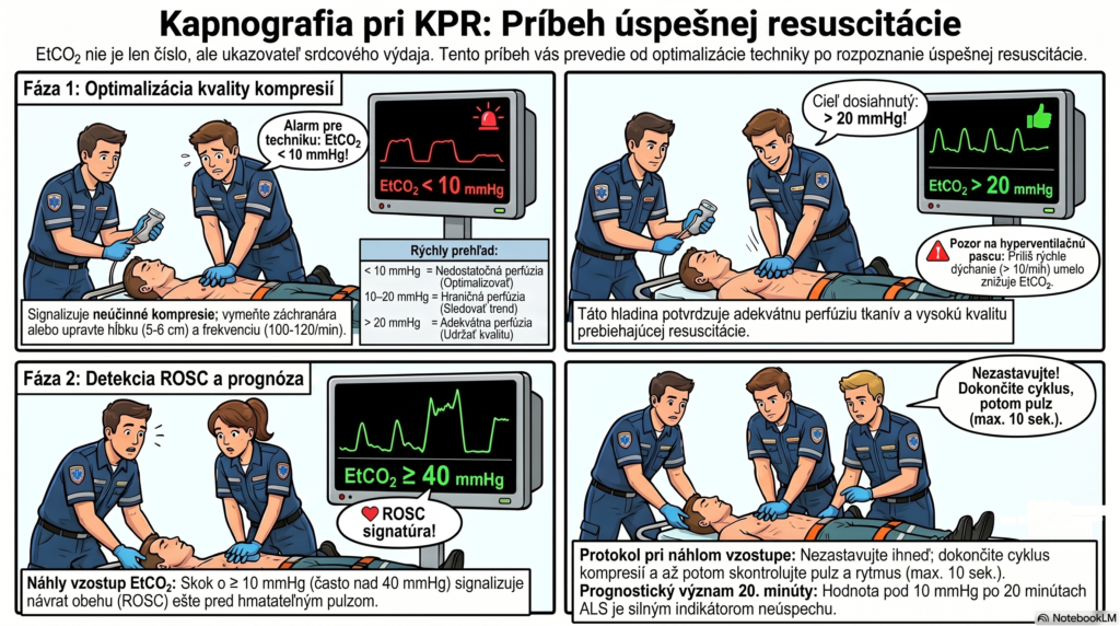
Capnography during CPR:
reading resuscitation quality in real time
EtCO₂ during cardiopulmonary resuscitation isn’t just another number on the monitor. It’s a direct, immediate, and continuous assessment of how effectively your crew is pumping blood to the patient’s brain.
Cardiac arrest is a situation where every second decides the patient’s neurological outcome. And yet — most crews watch the ECG and SpO₂ during CPR while the capnograph runs alongside, unnoticed. This article shows you why EtCO₂ is the most information-rich parameter available to you during resuscitation.
Why CO₂ is generated during CPR at all
The physiological basis is simple, yet important: CO₂ reaches the lungs and is exhaled only if blood is circulating. Cells continuously produce CO₂ — during cardiac arrest, this CO₂ accumulates in the tissues and venous blood, but doesn’t reach the lungs, because the heart isn’t pumping.
When we start CPR, external chest compressions substitute for the heart’s function. Some of the CO₂ accumulated in the tissues begins to be transported to the lungs — and this is exactly the amount of CO₂ the capnograph measures. The EtCO₂ level during CPR is thus a direct indicator of the cardiac output generated by compressions.
EtCO₂ during CPR = f (CO₂ production × cardiac output from compressions × ventilation)
CO₂ production is relatively constant in the first minutes of resuscitation. We control ventilation ourselves. The variable that primarily determines EtCO₂ during CPR is therefore cardiac output from compressions. A low EtCO₂ = weak compressions or another cause of low perfusion.
Four clinical roles of EtCO₂ during CPR
The European Resuscitation Council (ERC 2021) recommends continuous capnographic monitoring in intubated patients during CPR for four clearly defined purposes. Let’s look at each in detail.
Role 1 — Immediate feedback on compression quality
This is the most practical function of capnography during resuscitation. EtCO₂ tells you in real time whether you’re compressing well — without needing to interrupt CPR to check a pulse, without subjective judgment.
| EtCO₂ during compressions | Interpretation | Crew action |
|---|---|---|
| < 10 mmHg | Inadequate perfusion — compressions are ineffective or there’s another cause | Check technique: depth (5–6 cm), rate (100–120/min), recoil. Rule out tension pneumothorax, tamponade. Swap the paramedic doing compressions. |
| 10–20 mmHg | Borderline perfusion — optimize | Improve technique, watch the trend. Consider mechanical CPR (LUCAS) if available. |
| > 20 mmHg | Adequate perfusion — good CPR quality | Maintain quality, watch for a further rise as a ROSC signal. |
After 4 minutes of compressions, EtCO₂ drops to 11 mmHg. The driver reports the paramedic doing compressions looks tired. Without capnography, no one would have noticed the drop in quality — the paramedic would have continued under the illusion that CPR was going well.
Swapping the paramedic on compressions → within 30 seconds EtCO₂ rises back to 20 mmHg. The capnograph just saved the quality of the resuscitation.
Role 2 — Detecting ROSC without interrupting compressions
This may be the most dramatic clinical application of capnography during CPR. Return of spontaneous circulation (ROSC) causes the heart to start pumping blood again — including the large amount of CO₂ that accumulated during the arrest. The result is a sudden, marked rise in EtCO₂ — typically from 15–25 mmHg during CPR to 40–60 mmHg or more.
According to ERC 2021, a rapid rise in EtCO₂ during CPR precedes a palpable pulse by several minutes. This means the capnograph detects ROSC before the paramedic manages to palpate it — letting them skip the interruption of compressions to check a pulse exactly at the most critical moment.
1. EtCO₂ suddenly rises ≥ 10 mmHg or exceeds 40 mmHg → finish the current compression cycle, don’t stop immediately
2. At the end of the cycle → pause to check rhythm and pulse (max. 10 seconds)
3. If a pulse is palpable + an organized rhythm → confirm ROSC, begin post-resuscitation management
4. If no pulse is palpable → continue CPR, watch whether EtCO₂ remains elevated (it may be a movement artifact from a crew swap)
Role 3 — Monitoring ventilation during CPR
The hyperventilation trap during CPR is a real problem. Under the pressure of the situation, paramedics naturally tend to ventilate the patient too fast — increased intrathoracic pressure reduces venous return, paradoxically worsening cardiac output from compressions.
ERC 2021 recommends ventilating an intubated patient during CPR at a rate of 10/min — one breath every 6 seconds. The capnograph displays the number of waveforms per minute, allowing precise adherence to this rate without needing to count.
Ventilating too fast (RR > 12/min in an intubated patient) increases intrathoracic pressure, reduces venous return to the right heart, and thereby reduces compression effectiveness — the exact opposite of what the paramedic intends.
EtCO₂ during hyperventilated CPR can be paradoxically low not because compressions are poor, but because you’re exhaling CO₂ too fast. Watch the number of waveforms on the monitor — not the feeling that you’re ventilating enough.
Role 4 — A prognostic tool and decision support for terminating CPR
This is the clinically and ethically most sensitive role of capnography during resuscitation. ERC 2021 explicitly states that EtCO₂ must never be used as the sole factor in deciding to terminate CPR — it’s always a combination of multiple criteria. Nevertheless, it plays an important role in the overall clinical picture.
Key studies — including Levine’s 1997 paper in the New England Journal of Medicine and its later re-analyses — show that an EtCO₂ < 10 mmHg after 20 minutes of quality ALS resuscitation in an intubated patient with a non-shockable rhythm is a strong predictor of mortality. More recent data (Portland Cardiac Arrest Registry 2018–2021) suggest that tracking the EtCO₂ trend over time (delta-EtCO₂) has better discriminative value than a single reading.
EtCO₂ and reversible causes of cardiac arrest (4Hs/4Ts)
Capnography can also help with the differential diagnosis of the cause of cardiac arrest — not definitively, but as another clinical clue:
| Cause of arrest | Typical EtCO₂ pattern | Clinical context |
|---|---|---|
| Hypovolemia | Low, progressively declining | Trauma, GI bleeding — little blood = little CO₂ transported to the lungs |
| Tension pneumothorax | Sudden drop | Venous return obstruction → drop in cardiac output → drop in EtCO₂ |
| Cardiac tamponade | Low, unresponsive to compressions | The heart can’t fill → compressions ineffective → low EtCO₂ |
| Massive PE | Very low | Obstruction of the pulmonary vascular bed → CO₂ doesn’t reach the alveoli despite circulation |
| Hyperkalemia / intoxication | Variable | Depends on cardiac output — EtCO₂ helps track response to the antidote |
A practical capnography protocol on a cardiac arrest call — step by step
The capnograph isn’t relevant yet — without securing the airway, meaningfully measuring EtCO₂ isn’t possible. Priority: compressions, defibrillation.
The first capnographic waveform after intubation = confirmation of correct tube placement. Record the initial EtCO₂ value.
Goal: EtCO₂ > 20 mmHg. If below 10 mmHg → immediately optimize technique, swap the paramedic, rule out mechanical causes (tension pneumothorax).
During the pause: if EtCO₂ stays constant or rises → could be early ROSC. If it drops at every pause → continue CPR.
EtCO₂ < 10 mmHg + non-shockable rhythm + no reversible causes + no ROSC → discuss terminating resuscitation per the current EMS protocol and dispatch center guidance.
Finish the cycle → pulse + rhythm → if ROSC: target temperature, O₂ titration, SpO₂ 94–98%, transport to a PCI center / emergency department.
What EtCO₂ during CPR does not replace
In closing, an important note of clinical humility: capnography is a powerful tool, but it has limits the paramedic must know:
Epinephrine lowers EtCO₂. Giving epinephrine causes a transient EtCO₂ drop due to vasoconstriction and blood redistribution — don’t interpret a post-epinephrine drop as worsening compressions. Wait 2–3 minutes before evaluating.
Bicarbonate raises EtCO₂. NaHCO₃ releases CO₂ through a chemical reaction — a sudden EtCO₂ rise after giving bicarbonate isn’t ROSC. It’s an artifact.
EtCO₂ doesn’t replace clinical judgment. Pulse, rhythm, cause of arrest, patient context — all of it must be part of the decision-making process. EtCO₂ is one factor, not the only one.
- Capnography in EMS: why it’s your second monitor
- The capnographic waveform: reading the patient’s story
- Capnography during CPR: reading resuscitation quality in real time (this article)
- Capnography traps: when the waveform lies — esophageal intubation, bronchospasm, artifact
- EtCO₂ and the decision to terminate CPR: when to continue and when not to
- Capnography in the spontaneously breathing patient — opioid intoxication, status epilepticus, DKA
- Perkins GD, et al. European Resuscitation Council Guidelines 2021: Adult Advanced Life Support. Resuscitation. 2021;161:115–151. doi:10.1016/j.resuscitation.2021.02.010
- Levine RL, et al. End-Tidal Carbon Dioxide and Outcome of Out-of-Hospital Cardiac Arrest. N Engl J Med. 1997;337(5):301–306.
- Hartmann SM, et al. Utility of End-Tidal Carbon Dioxide to Guide Resuscitation Termination in Prolonged Out-of-Hospital Cardiac Arrest. Am J Emerg Med. 2023;75:32–37.
- Sheak KR, et al. Quantitative Relationship Between End-Tidal Carbon Dioxide and CPR Quality During Both In-Hospital and Out-of-Hospital Cardiac Arrest. Resuscitation. 2015;89:149–154.
- Savastano S, et al. Capnography During Cardiac Arrest. Resuscitation. 2017;120:1–5. doi:10.1016/j.resuscitation.2017.08.218
- EMS1. Capnography During CPR: 5 Things to Know About ETCO2 and Waveform Monitoring. ems1.com. April 2025.
- JEMS. The Role of EtCO2 in Termination of Resuscitation. jems.com. October 2024.
- ALiEM. End-Tidal CO2 in Cardiopulmonary Resuscitation. aliem.com. 2019.
- PMC / NCT. End-Tidal Carbon Dioxide Trajectory-Based Prognostication of Out-of-Hospital Cardiac Arrest. PMC11254150. 2024.
Paramedic with more than 15 years of experience in prehospital emergency care, RZP Leopoldov. Administrator and editor of zachranarjecool.eu. Author of the Capnography in EMS series.