
Neurologický deficit —ako vyšetrovaťod začiatku do konca

Neurological Deficit: How to Examine from Start to Finish

Zdieľajte
Assessing the Patient with a Neurological Deficit | zachranarjecool.eu
Patient Examination · VYS-NEU-01

Neurological Deficit —
how to examine
from start to finish

Step by step: how to distinguish stroke from hypoglycemia, how to correctly apply NIHSS in the field, what not to forget with an altered patient, and which signs are an immediate call for transport.

Time-critical Author: Martin Semanco, EMT-P Series: VYS-NEU-01 Reading time: ~13 min

Red Flags — immediate action

GCS ≤ 8 — airway management, intubation
Sudden headache — “worst headache of my life” — SAH?
Focal deficit + onset < 4.5 hrs — the thrombolysis window
Seizure lasting > 5 min or repeated without regaining consciousness
Anisocoria + altered consciousness — transtentorial herniation
BP > 220/120 + neurological deficit — hypertensive encephalopathy
Blood glucose < 3.5 mmol/l — measure this first, always
Trismus + opisthotonus + fever — meningitis/tetanus
⚠ Time is brain In stroke, approximately 1.9 million neurons die every minute without treatment. Time from onset to needle (door-to-needle) should be <60 minutes — your speed in the field directly affects how much brain the patient saves. Don’t waste time on unnecessary history-taking on scene — get the essentials, transport, and complete the rest en route.

History-taking — what and how to ask

With a neurological deficit, one single piece of data matters most: the exact time of symptom onset. Everything else is secondary. If the patient was asleep and woke up with a deficit — onset time = the time they were last seen without symptoms (last known well).

LetterTermHow to ask and what to concludeRed Flag answer
OOnset “When exactly did you first notice this symptom? What were you doing at that moment?”
Sudden onset during activity = hemorrhagic stroke or SAH. Gradual development over minutes–hours = ischemic stroke or TIA. Episodic = TIA, migraine, epilepsy.
Onset < 4.5 hrs from last known well = possible thrombolysis — pre-notify the hospital immediately
PProvocation “Did anything happen beforehand? Head injury, physical exertion, stress, alcohol, a medication?”
Head injury → epidural/subdural hematoma. Exertion + headache → SAH. Missed AED dose → seizure. Insulin without food → hypoglycemia.
Head injury + loss of consciousness + lucid interval → epidural hematoma — urgent transport
QQuality “What exactly do you feel — weakness, numbness, trouble speaking, double vision, dizziness?”
Weakness + speech + face = classic (cortical) stroke. Dizziness + double vision + ataxia = posterior circulation (vertebrobasilar). Isolated sensory deficit without motor involvement = TIA or lacunar stroke.
Paralysis + speech + face all at once = a large cortical stroke, possible LVO — consider a thrombectomy center
RRadiation / distribution “Is it on the whole one side of the body, or just the arm? The face too? The leg too?”
Hemiplegia including the face = cortical lesion (MCA). Only arm and face without the leg = cortical hand. Only the leg = ACA lesion. Bilateral = brainstem lesion or metabolic cause.
Bilateral deficit + altered consciousness = brainstem stroke or metabolic encephalopathy
SSeverity “Can the patient stand, walk, speak? Do they understand you? Is it improving or worsening since onset?”
Improving deficit = TIA or reperfusion. Worsening = stroke progression or bleeding. Fluctuating = a hemodynamic cause (stenosis, fibrillation).
Rapidly worsening deficit = malignant edema, hemorrhagic transformation — a neurosurgical center
TTime “When did someone last see you without any problems? When did you wake up? Do you have a watch that shows the time?”
Last known well is key for the thrombolysis decision. If the patient was found after sleeping — LKW = the time they went to sleep.
LKW < 4.5 hrs = pre-notify the stroke center, bypass a local hospital if thrombectomy is available
⚡ Additional mandatory questions Anticoagulants (warfarin, DOACs) — bleeding into the brain is higher risk and thrombolysis is contraindicated. Antiepileptics — a missed dose? Diabetes + insulin — hypoglycemia mimics stroke perfectly. Previous stroke or TIA — a recurrence? Migraine with aura — hemiplegic migraine mimics stroke.

Neurological examination — sequence and technique

In the field you perform a targeted, fast neurological exam — not a comprehensive neurology consult. The goal: detect the presence and extent of a deficit, estimate the location of the lesion, and decide on transport destination. Use a systematic approach.

Consciousness and GCS

The first step — it determines the urgency of everything else
Procedure — how to do it
1
AVPU — rapid orientation in the first seconds. Alert (responds spontaneously), Voice (responds when addressed), Pain (responds only to pain), Unresponsive (no response). AVPU gives you immediate information even before GCS. If the patient is V or P — GCS will be ≤13; if U — GCS ≤8. Think airway management.
2
GCS — three components separately, not the total. Eyes (E 1–4): spontaneous, to voice, to pain, none. Verbal (V 1–5): oriented, confused, inappropriate words, sounds, none. Motor (M 1–6): follows command, localizes, withdraws, flexion, extension, none. Always document as “E3V4M5 = GCS 12,” not just the number. The GCS trend (improving/worsening) matters more than one reading.
3
Painful stimulus — correct technique. Use a central stimulus (rubbing the sternal notch with your knuckles) or peripheral (nail bed pressure). Central assesses consciousness, peripheral assesses limb motor response — use both. Never pinch the skin — it’s not a standardized stimulus and doesn’t reliably score GCS.
4
Orientation — if the patient is conscious. Person (do they know who they are?), place (where are we?), time (date, year, day). Situation (what happened?). Disorientation to time appears first — before disorientation to person. Don’t assess orientation just from whether the patient “looks fine.” Ask directly.
What you find and how to assess it

Normal finding

  • GCS 15, fully oriented
  • Spontaneously open eyes, appropriate contact
  • Responds correctly to questions

Pathological finding

  • GCS ≤ 8 — secure the airway, intubation
  • Rapid GCS decline — herniation, bleeding, edema
  • Disorientation — stroke, metabolic encephalopathy, intoxication
  • Flexion/extension rigidity — deep coma, very poor prognosis

Speech and language

Aphasia vs. dysarthria — two different lesions, two different approaches
Procedure — how to do it
1
Name objects. Show the patient a pen, a watch, a mobile phone. Ask them to tell you what it is. Anomia (can’t name it) = aphasia. Names it correctly but unclearly = dysarthria. Aphasia = a language disorder (comprehension or production). Dysarthria = a motor speech disorder — the patient knows what they want to say, they just can’t articulate it. Clinically completely different.
2
Comprehension — a simple command. Ask the patient: “Raise your right hand.” Then: “Show me your left ear.” If they don’t understand = a comprehension disorder (Wernicke’s aphasia or global aphasia). A patient with Wernicke’s aphasia speaks fluently but without meaning — sounds like confused speech. They’re often unaware of the deficit themselves. Don’t mistake this for intoxication.
3
Sentence repetition. Ask the patient to repeat: “Today is a nice day.” Inability to repeat with preserved comprehension = conduction aphasia (arcuate fasciculus lesion). In the field, it’s enough to distinguish: speaks/doesn’t speak, understands/doesn’t understand. Precise aphasia classification is a neurologist’s task.
What you find and how to assess it

Normal finding

  • Fluent, intelligible speech
  • Correctly names objects
  • Understands commands, repeats sentences

Pathological finding

  • Aphasia — a cortical lesion of the left (dominant) hemisphere
  • Global aphasia — neither speaks nor understands, a large MCA lesion
  • Dysarthria — brainstem lesion, cerebellar stroke, intoxication
  • Normal speech doesn’t rule out stroke — a silent lesion is possible!

Motor and sensory function

Test both sides deliberately — asymmetry is key
Procedure — how to do it
1
Motor drift — the Mingazzini test for the arms. Ask the patient to extend both arms forward with palms up and close their eyes. Normal: both arms hold position for 10 seconds. Pathological: one arm drops, pronates, or drifts — that’s paresis, even if not obvious. Drift is more sensitive than gross strength testing. It catches even mild paresis the patient consciously compensates for.
2
Motor drift — legs. Patient supine, raises both legs to 30° and holds for 5 seconds. One leg dropping = paresis. Alternative: ask the patient to press hard against your hands with both feet — compare the strength on each side. For a supine patient, don’t forget to test the legs — posterior-circulation stroke can present with only a leg motor deficit without arm involvement.
3
Grip strength — both hands at once. Ask the patient to squeeze your fingers with both hands simultaneously. Compare sides. Grip asymmetry = a contralateral hemispheric lesion. Don’t assess strength just verbally — “do you feel weak?” is unreliable. Physically measure and compare sides.
4
Sensation — comparing sides. Prick the skin with a sterile needle or pinch the skin — compare the right and left side of the face, arm, leg. The patient tells you whether they feel it equally on both sides. Hemihypesthesia (reduced sensation on one side) + hemiparesis = a thalamic lesion or cortical stroke.
Specific tests — how they’re performed

FAST score +

Rapid prehospital stroke detection.

Face: Ask them to smile — facial asymmetry? Arms: Mingazzini — one arm drops? Speech: Repeat a sentence — aphasia or dysarthria? Time: Record the onset. If one of the first three is positive = high probability of stroke.

Cincinnati Stroke Scale

An alternative to FAST — the same three items, statistically validated.

Facial asymmetry + arm drift + speech. Sensitivity for stroke 66%, specificity 87%. Positive predictive value with one sign 72%. One positive finding is enough to activate the stroke protocol.

Abbreviated (field) NIHSS

Orientation-level neurological scoring in the field.

Assess: consciousness (0–3), gaze (0–2), visual field (0–3), face (0–3), arm motor L+R (0–4 each), leg motor L+R (0–4 each), speech (0–3), language/dysarthria (0–3). Score >15 = a large stroke, consider a thrombectomy center.

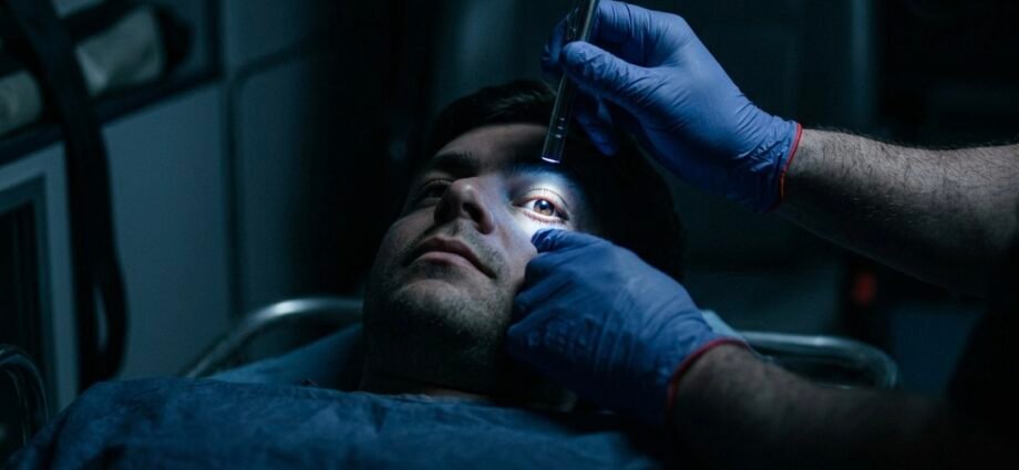
Pupillary reflexes

Assessing brainstem function and herniation.

A flashlight from the side — direct reflex (constriction of the illuminated eye) and indirect/consensual (constriction of the other eye). Anisocoria (a difference >1 mm) + altered consciousness = transtentorial herniation on the side of the larger pupil. Immediate hyperventilation + transport.

Romberg test

Assessing sensory ataxia — if the patient is standing.

Patient stands, feet together, arms at their sides. They close their eyes. Positive = marked swaying or falling when the eyes are closed. Cerebellar ataxia is positive even with eyes open. Romberg assesses sensory pathways (proprioception), not the cerebellum.

Finger-to-nose test

Assessing cerebellar function.

The patient alternately taps their finger to the tip of their nose and to your finger, which you move. Positive = dysmetria (misses the target) or intention tremor (trembles as it approaches the target). An ipsilateral cerebellar lesion — cerebellar stroke.
ℹ Clinical pearl — cerebellar stroke Cerebellar stroke presents with dizziness, ataxia, and vomiting — and is easily mistaken for labyrinthitis or BPPV. The key difference: the HINTS test. Head Impulse (normal = a central lesion!), Nystagmus (direction-changing = central), Test of Skew (vertical deviation = central). Untreated cerebellar stroke can cause fatal cerebellar edema within 24–48 hours.

Eyes and cranial nerves

Pupils, gaze, facial expression — localizing information
Procedure — how to do it
1
Pupils — size, symmetry, reactivity. Normal: 3–5 mm, isocoric, rapid light response. Assessed in dim light, not direct sunlight. Check for medications that alter pupils (opioids = miosis, anticholinergics = mydriasis). Unilateral fixed mydriasis + altered consciousness = transtentorial herniation on that same side. Bilateral fixed mydriasis = deep coma, herniation, or intoxication (atropine, cocaine).
2
Conjugate gaze — eye deviation. Ask the patient to follow your finger side to side and up/down. Normal: both eyes move synchronously. Deviation of both eyes to one side = “the patient looks toward their lesion” (cortical) or “looks away from the lesion” (brainstem). Cortical lesion: eyes deviate ipsilateral to the lesion (away from the paresis). Brainstem (pontine) lesion: eyes deviate contralateral to the lesion (toward the paresis). Distinguishing this helps localize the lesion.
3
Facial symmetry — CN VII. Ask the patient to smile, show their teeth, frown, puff out their cheeks. Asymmetry = CN VII paresis. Key: central (stroke) vs. peripheral (Bell’s palsy) CN VII paresis. Central paresis: the lower third of the face is affected contralaterally, the forehead is spared (the patient can still frown). Peripheral paresis (Bell’s): the whole ipsilateral face, forehead included. In the field: if they can’t frown = peripheral, not stroke.
4
Nystagmus — if there are complaints of dizziness. Ask the patient to follow your finger — moving up to 30° from center. Nystagmus: a fast and a slow phase. Unilateral horizontal = peripheral (labyrinthine). Vertical or direction-changing = central (brainstem or cerebellum), urgent. Direction-changing (gaze-evoked) nystagmus = always central = don’t rule out stroke. Fixed unilateral horizontal nystagmus = likely a peripheral vestibular problem.
What you find and how to assess it

Normal finding

  • Isocoric pupils 3–5 mm, rapid reflex
  • Synchronous conjugate gaze
  • Symmetric facial expression, forehead symmetric
  • No nystagmus on gaze to 30°

Pathological finding

  • Anisocoria + altered consciousness — herniation, immediately
  • Bilateral fixed mydriasis — deep coma, extremely poor prognosis
  • Conjugate eye deviation — a cortical or brainstem lesion
  • Vertical nystagmus — a brainstem lesion, central pathology

Vital signs and supplementary measurements

The minimum standard — without exception
What to measure, why, and how to interpret it
1
Blood glucose — always first, without exception. Hypoglycemia (glucose <3.5 mmol/l) causes a focal neurological deficit, hemiplegia, speech disturbance, altered consciousness — indistinguishable from stroke without a glucometer. Treating hypoglycemia is simple and immediately reversible. Don’t treat as stroke until you’ve ruled out hypoglycemia. A paramedic who doesn’t check blood glucose in a patient with a neurological deficit and administers stroke treatment is making a fundamental error. This is one of the most common causes of complaints and malpractice disputes.
2
BP — both arms if dissection is suspected. Hypertensive encephalopathy: BP >220/120 + a neurological deficit. A BP difference between arms >20 mmHg = aortic dissection involving the branching cerebral arteries. With confirmed stroke: target BP before thrombolysis <185/110. Aggressive antihypertensive treatment in the field is contraindicated — the brain in acute ischemia needs a higher BP to perfuse the penumbra.
3
12-lead ECG. Atrial fibrillation causes ~30% of ischemic strokes (cardioembolic etiology). New AV blocks and Q waves can indicate cardiogenic embolism. Ventricular tachycardia and other arrhythmias can cause syncope mimicking stroke. Don’t delay the ECG — run it in parallel with history-taking, not only after the physical exam.
4
Temperature. Fever + neck stiffness + altered mental status = meningitis/encephalitis — isolation, pre-notification. Hyperpyrexia (T >40°C) alone can cause encephalopathy and seizures. Sepsis with encephalopathy mimics stroke in elderly patients. Neck stiffness: patient supine, passively flex the head toward the chest. If they resist and complain of pain = meningismus = urgent pre-notification.
5
SpO₂ and breathing. Hypoxia (SpO₂ <94%) worsens ischemic stroke — give O₂. Cheyne-Stokes breathing = bilateral hemispheric dysfunction. Ataxic breathing = a brainstem lesion, pre-arrest. Kussmaul breathing = metabolic acidosis (not neurological). Hyperventilation in the field is indicated only for known herniation (anisocoria, extensor rigidity) — otherwise it worsens the stroke outcome through vasoconstriction.

Differential diagnosis — stroke mimics and true stroke

Not every neurological deficit is a stroke. Up to 20–30% of patients transported as “stroke” have another cause — so-called stroke mimics. Correctly distinguishing them determines transport destination and prevents unnecessary thrombolysis.

DiagnosisKey signs and distinguishing featuresUrgency
Ischemic stroke — LVOAphasia + hemiplegia + eye deviation, NIHSS >10, onset <24 hrs, AFib historyLIFE-THREAT
Hemorrhagic strokeSudden onset, headache, vomiting, rapid progression, BP ↑↑, anticoagulantsLIFE-THREAT
SAH — subarachnoid hemorrhage“Worst headache ever” — onset in seconds, meningismus, photophobia, vomitingLIFE-THREAT
Epidural hematomaHead injury + a lucid interval + rapid GCS deterioration, ipsilateral anisocoriaCRITICAL
Status epilepticusSeizure >5 min or a series without regaining consciousness, cyanosis, incontinence, tongue bitingCRITICAL
HypoglycemiaGlucose <3.5 mmol/l, diabetes, insulin, a focal deficit — responds to glucoseURGENT
Hypertensive encephalopathyBP >220/120, headache, confusion, visual disturbance, no focal deficitURGENT
Meningitis / encephalitisFever + neck stiffness + photophobia + altered status, Kernig/Brudzinski +URGENT
Todd’s paresis (post-ictal)After a seizure — hemispheric paresis lasting min–hrs, resolves spontaneously, epilepsy historyURGENT
TIA — transient ischemic attackFocal deficit <24 hrs (typically <1 hr), full resolution, ABCD² scoreURGENT
Hemiplegic migraineMigraine history, aura, headache following the deficit, young patient, family historySEMI-URGENT
Conversion disorderYoung patient, psychological stress, an inconsistent finding, preserved strength with distractionSEMI-URGENT
ℹ The rule for uncertainty If you can’t reliably distinguish stroke from a stroke mimic — transport as stroke. The cost of a false-positive stroke protocol activation is much lower than the cost of a missed window for thrombolysis or thrombectomy. In dubio pro stroke.

Sources used

The content of this article is based on current guidelines and professional textbooks of emergency and neurological medicine.

01
ESO Guidelines for the Management of Ischaemic Stroke, 2021
Berge E, et al. European Stroke Organisation. European Stroke Journal, 2021. DOI: 10.1177/23969873211012177
Diagnostic criteria for ischemic stroke, thrombolysis indications (the 4.5-hr window), thrombectomy indications, prehospital management.
02
AHA/ASA Guidelines for the Early Management of Patients with Acute Ischemic Stroke, 2019
Powers WJ, et al. Stroke, 2019. DOI: 10.1161/STR.0000000000000211
NIHSS scoring, BP management before thrombolysis, prehospital pre-notification, bypass protocols for stroke centers.
03
Tintinalli’s Emergency Medicine: A Comprehensive Study Guide, 9th ed.
Tintinalli JE, et al. McGraw-Hill Education, 2019.
Physical neurological examination, differential diagnosis of stroke mimics, GCS, pupillary reflexes, aphasia vs. dysarthria.
04
Pre-Hospital Stroke Scales in Clinical Practice: A Systematic Review
Zhelev Z, et al. BMJ Open, 2019. DOI: 10.1136/bmjopen-2018-023379
Validation of the FAST score and Cincinnati Stroke Scale in the prehospital setting, sensitivity and specificity, scale comparison.
05
HINTS to Diagnose Stroke in the Acute Vestibular Syndrome
Kattah JC, et al. Stroke, 2009. DOI: 10.1161/STROKEAHA.109.551234
The HINTS test (Head Impulse, Nystagmus, Test of Skew) for differentiating peripheral vs. central vertigo syndrome, cerebellar stroke.
06
Urgentní medicína v klinické praxi lékaře
Šeblová J, Knor J et al. Grada Publishing, 2013. ISBN 978-80-247-4434-6.
The prehospital approach to stroke, seizures, and neurological emergencies within the Slovak/Czech EMS system.
07
Slovak Act No. 579/2004 Coll. on Emergency Medical Services
National Council of the Slovak Republic. Collection of Laws of the Slovak Republic.
Scope of a paramedic’s competency for neurological examination, glucose administration, and stroke-center pre-notification.
✓ Article update Content reflects the current ESO Guidelines 2021 and AHA/ASA 2019. The article will be revised if guidelines or legislation change. Report inaccuracies via the contact form on zachranarjecool.eu.

Leave a Reply

Your email address will not be published. Required fields are marked *