ABSTRACT
Interpreting the electrocardiogram (ECG) in prehospital emergency medical care is a critical paramedic competency. Given the high level of stress factors and time pressure, frequent diagnostic errors occur in the field due to an unsystematic approach. The RAFTING system is a methodical, cognitive framework that forces the examiner into sequential evaluation of the electrical events in the myocardium. This article analyzes in detail each component of the algorithm, from basic rhythm analysis to advanced QT-interval measurement, and their direct application in paramedic clinical practice.
1. INTRODUCTION
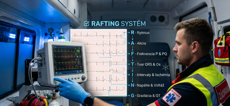
In modern emergency medicine, diagnosing acute conditions relies on rapid synthesis of clinical symptoms and supplementary tests. In prehospital care, the ECG trace is a key determinant of treatment strategy, particularly when deciding whether to activate an interventional cardiac center or administer specific antiarrhythmic treatment. A persistent problem in practice is so-called “selective perception,” where the paramedic subconsciously looks only for signs of acute myocardial infarction (STEMI), causing other, often fatal, changes to be overlooked. Implementing the RAFTING mnemonic (Rhythm, Action, Frequency, Type, Intervals, Numbers, General impression) serves as a safety protocol that ensures a comprehensive, objective description of the trace regardless of whether a dominant pathology is present.
2. CORE: COMPREHENSIVE ANALYSIS OF THE RAFTING SYSTEM PARAMETERS
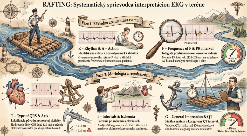
R – Rhythm
The initial analysis must reliably identify the heart’s primary pacemaker and the regularity of impulse generation. Physiological sinus rhythm is defined not only by the presence of a P wave before each QRS complex, but also by its constant morphology and axis, which must be positive in lead II and negative in aVR. At this stage, the paramedic distinguishes between a regular rhythm and irregularities that may result from respiratory arrhythmia (benign) or more serious conditions such as atrial fibrillation or junctional escape rhythms. Understanding the rhythm’s origin mechanism is fundamental to distinguishing supraventricular from ventricular disorders.
A – Action (Rate)
Determining heart rate in prehospital care goes beyond simply counting beats per minute. The paramedic must account for the paper speed (standardly 25 mm/s), using the mathematical ratio (300/number of large squares between R waves) for a regular rhythm. A critical aspect, however, is clinically interpreting the rate in the context of hemodynamic stability; tachycardia above 150 bpm or bradycardia below 40 bpm requires immediate intervention per ALS protocols. Correctly determining the rate is also essential for the later calculation of the corrected QT interval.
F – Frequency of P & PR interval (Atrial Activity and Conduction)
This part of the algorithm focuses on the integrity of atrioventricular conduction and the morphology of atrial depolarization. The PR interval, with a physiological duration of 120 to 200 ms, reflects the time needed for the impulse to pass through the AV node. Prolongation of this interval beyond 200 ms diagnoses 1st-degree AV block, while progressive lengthening or sudden dropped beats signal higher-degree blocks (Wenckebach, Mobitz II), which can result in an asystolic pause in the field. P-wave morphology also provides indirect information about pulmonary circulation pressure (P-pulmonale) or left atrial dilation (P-mitrale).
T – Type of QRS & Axis (QRS Morphology and Electrical Axis)
QRS complex analysis is key for localizing the origin of ventricular activity. A complex width under 110 ms indicates rapid impulse propagation through the His-Purkinje system, while widening beyond 120 ms signals aberration, bundle branch block (LBBB/RBBB), or an ectopic ventricular focus. It’s also necessary to determine the heart’s electrical axis in the frontal plane using the quadrant method (leads I and aVF). Pathological left axis deviation can indicate left anterior hemiblock (LAH), while right axis deviation with acute dyspnea raises suspicion of acute right ventricular strain, for example in massive pulmonary embolism.
I – Intervals & Ischemia (ST Segment and T Wave)
The repolarization phase is the ECG segment most commonly scrutinized in emergency medicine for diagnosing acute coronary syndrome (ACS). The paramedic assesses the ST segment’s position relative to the isoelectric line (measured at the TP segment), looking for J-point elevations that define STEMI, or depressions signaling subendocardial ischemia. The T wave must be interpreted in the context of the dynamics of change; tall, symmetric, peaked T waves are often the first marker of the hyperacute phase of infarction or of severe hyperkalemia, which in the prehospital setting poses an immediate threat to the patient’s life.
N – Numbers & Voltage (Amplitude and Voltage)
Quantitative analysis of deflections provides information about the myocardium’s electrical strength and the conductivity of the surrounding environment. High-amplitude R waves in the chest leads, meeting Sokolow-Lyon index criteria, point to chronic left ventricular hypertrophy, often associated with arterial hypertension. Conversely, diffusely low voltage (deflections under 0.5 mV in the limb leads) can, in prehospital practice, warn of pericardial effusion with cardiac tamponade risk, or of tension pneumothorax, where the air layer acts as an insulator.
G – General Impression & QT (Overall Impression and Corrected QT Interval)
The algorithm’s final step serves to verify parameters that escape primary attention. The most critical is measuring the QT interval, which must be corrected for heart rate (QTc). QTc prolongation beyond 470 ms represents a substrate for the malignant ventricular arrhythmia Torsades de Pointes. The general impression closes the analysis by synthesizing all findings into a final working diagnosis, also taking into account the presence of pathological U waves or other artifacts that could lead to misinterpretation of the trace.
3. DISCUSSION
Scientific studies in cognitive psychology confirm that structured checklists and mnemonics dramatically reduce error rates in medicine. The RAFTING system transfers this principle to ECG interpretation in prehospital emergency medical care. Its value rests on the fact that while an experienced physician can evaluate a trace intuitively (pattern recognition), for a paramedic and a student a systematic approach is the only guarantee of patient safety. The algorithm eliminates the risk of missing “silent” killers such as a prolonged QT interval or ECG signs of pulmonary embolism, which are not as obvious as massive ST elevations.
4. CONCLUSION
Systematic ECG description using the RAFTING system is a standardized method that increases diagnostic accuracy in emergency medicine. Implementing this algorithm in paramedic education in Slovakia fully aligns with requirements for improving the quality of healthcare provided. Systematizing interpretation is not merely a formal requirement, but a necessary precondition for reducing mortality and morbidity in the prehospital management of critical conditions.
5. REFERENCES
- DOBIÁŠ, V. et al. (2021). Prehospital Emergency Medicine. 3rd edition. Martin: Osveta.
- HAMPTON, J. R. (2019). The ECG Made Easy. 9th edition. Elsevier.
- ŠANDRIKOVÁ, V. (2018). Electrocardiography for Non-Physician Healthcare Professions.
- Guidelines of the European Society of Cardiology (ESC) for the management of acute coronary syndromes.
- Slovak Government Regulation No. 296/2010 Coll. on professional competence for the practice of healthcare professions.