ABSTRACT
The second step of the RAFTING diagnostic algorithm, represented by the letter “A” (Action / Rate), defines the procedures for precisely determining heart rate (HR) in prehospital emergency medical care. This article analyzes the mathematical models for calculating rate at standard paper speed and focuses primarily on the clinical interpretation of the obtained value in the context of the patient’s hemodynamic stability. Correctly assessing the rate is not only a precondition for selecting the appropriate branch of European Resuscitation Council (ERC) algorithms, but also a necessary input value for calculating the corrected QT interval (QTc).
1. INTRODUCTION
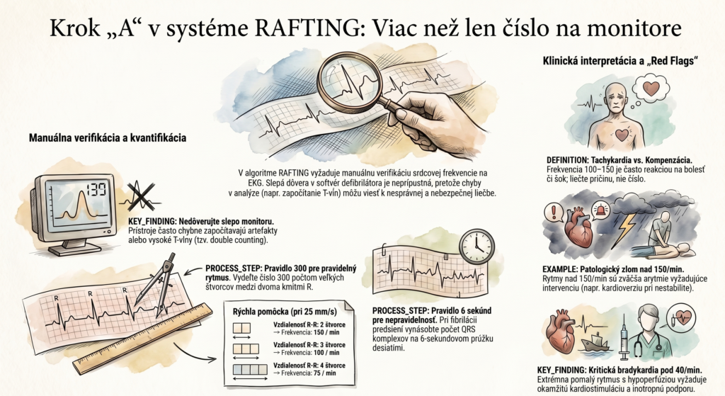
In the era of modern digital defibrillators that automatically generate a numeric heart-rate value on the display, manually assessing rate may seem redundant. For a professional paramedic, however, blind trust in the device’s software analysis is unacceptable. Devices are often prone to errors (so-called double counting of tall T waves or artifacts), which can lead to incorrect indication of antiarrhythmic treatment. Step “A” in the RAFTING system therefore forces the examiner to verify the hardware data and to understand that heart rate isn’t just an isolated number, but a key determinant of cardiac output (CO = HR x SV) and myocardial oxygen consumption.
2. CORE: METHODOLOGY AND CLINICAL CORRELATION
2.1 Manual Rate Quantification
The basic precondition for a successful calculation is knowing the paper speed, which in the European and Slovak standard is set primarily to 25 mm/s. At this speed, one large square (5 mm) corresponds to a time interval of 0.20 s, and one second represents 5 large squares. The paramedic uses two basic methods:
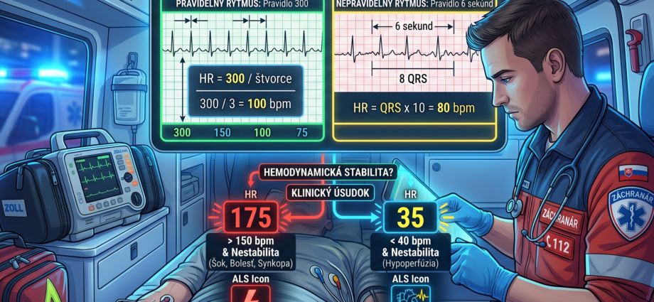
- Regular rhythm (the 300 rule): Based on the fact that the device prints 300 large squares per minute (60 s / 0.20 s). Heart rate is calculated as: 300 / number of large squares between two consecutive R waves. (E.g., a distance of 3 squares = a rate of 100/min). For maximum precision with fast rhythms, the 1500 rule is used (1500 / number of small squares).
- Irregular rhythm (the 6-second rule): With atrial fibrillation, the 300 rule is unusable. The paramedic counts the number of QRS complexes on a 6-second strip (30 large squares) and multiplies it by 10.
2.2 Clinical Interpretation and Differential Diagnosis
The rate value directly shapes the paramedic’s diagnostic view and opens specific differential-diagnostic windows:
- Rate 100–150/min (tachycardia): This is most often sinus tachycardia, usually a compensatory mechanism. Here, the paramedic must not primarily treat the rate itself (e.g., by giving beta-blockers), but must find and remove the cause — hypovolemia (hemorrhage, dehydration), sepsis, hypoxia, pain, or intoxication.
- Rate > 150/min: At these values, the physiological SA node rarely takes control (except during extreme exertion). It’s almost always a pathological arrhythmia (SVT, atrial flutter with 2:1 conduction, or VT). At extreme rates, diastolic time shortens dramatically, leading to reduced ventricular filling and critical subendocardial ischemia.
- Rate < 60/min (bradycardia): While this reflects vagal tone in athletes, in a symptomatic patient in prehospital care we must consider conduction-system failure (AV blocks), ischemia (particularly RCA occlusion affecting the AV node), or iatrogenic overdose (beta-blockers, calcium channel blockers).
2.3 Downstream Relevance: Significance for QTc
Heart rate is absolutely essential for assessing the QT interval in the later “G” step of the RAFTING system. Since the QT interval physiologically shortens with tachycardia and lengthens with bradycardia, it must be recalculated to a rate of 60 beats per minute using Bazett’s formula. Without a precise HR value, assessing Torsades de Pointes risk is impossible.
3. RED FLAGS IN PREHOSPITAL PRACTICE
In the context of step “A,” the paramedic must immediately sharpen attention and prepare for radical intervention per Advanced Life Support (ALS) procedures in two borderline situations:
- Rate > 150/min with instability: If tachycardia is accompanied by signs of hemodynamic instability (shock state, syncope, myocardial ischemia, or acute heart failure), pharmacotherapy takes a back seat, and the primary choice is synchronized electrical cardioversion.
- Rate < 40/min with instability: Extreme bradycardia with hypoperfusion that doesn’t respond to atropine (or where atropine is contraindicated, e.g., in a denervated transplanted heart or with infranodal blocks) requires immediate initiation of transcutaneous pacing (TCP) and inotropic support (epinephrine/dopamine).
- A rate machine-like constant at 150/min: This specific finding (particularly with a narrow QRS) is a classic ECG “red flag” for atrial flutter with fixed 2:1 conduction. The atrial rate here is 300/min, and the AV node lets through every second impulse.
4. DISCUSSION
The biggest pitfall in paramedic practice is the phenomenon of “treat the monitor, not the patient.” Clinical practice shows cases where a device extrapolates a rate of 160/min because it mistakenly counts tall, peaked T waves (in hyperkalemia) as real QRS complexes, when the actual ventricular rate is 80/min. Manual verification of rate and correlating the ECG finding with palpation of the peripheral pulse (to rule out a pulse deficit in ectopy) are irreplaceable steps in safe patient management.
5. CONCLUSION
Quantifying heart rate within the RAFTING system is not merely a simple mathematical operation, but a deep clinical judgment. In a split second, the paramedic must assess whether the observed rate is the primary cause of the patient’s critical state (an arrhythmia requiring electrical or pharmacological intervention), or merely a physiological compensation for another underlying condition (bleeding, sepsis). Early recognition of borderline values and their hemodynamic consequences is a pillar of successful prehospital resuscitation and stabilization.
6. REFERENCES
- DOBIÁŠ, V. et al. (2021). Prehospital Emergency Medicine. 3rd edition. Martin: Osveta.
- European Resuscitation Council (ERC). (2021). Guidelines for Resuscitation: Adult advanced life support.
- HAMPTON, J. R. (2019). The ECG Made Easy. 9th edition. Elsevier.
- TÁBORSKÝ, M. et al. (2021). Clinical Cardiology. Mladá fronta.
- Slovak Ministry of Health guidance No. 11674/2010-OZSO on the provision of prehospital emergency medical care.