ABSTRACT
Identifying the underlying heart rhythm is the fundamental, first step (the letter “R” in the RAFTING algorithm) in ECG interpretation. This step requires reliably detecting the primary pacemaker and assessing the regularity of electrical impulse generation. This article analyzes in detail the electrophysiological criteria for physiological sinus rhythm, the differential diagnosis of irregularities, and the identification of escape or ectopic rhythms. The goal is to highlight diagnostic pitfalls and define red flags that are key for a prehospital paramedic in enabling early therapeutic intervention.
1. INTRODUCTION
Connecting a patient to a vital-signs monitor and obtaining a 12-lead ECG trace is standard for any patient with suspected cardiovascular failure. The first question a paramedic must ask before evaluating any other parameters (such as ST segments or QRS width) is: “Which anatomical focus is driving the heart’s electrical activity?” Step “R” (Rhythm) isn’t just about noting that activity is present, but about deeply understanding the hierarchy of the cardiac conduction system and the principle of overdrive suppression (suppression of lower-center activity by a faster dominant center).
2. CORE: RHYTHM DIAGNOSIS AND DIFFERENTIATION
2.1 Strict Criteria for Physiological Sinus Rhythm (SR)
The sinoatrial (SA) node is the primary physiological pacemaker. In emergency medicine, noting the “presence of any P wave” is not sufficient to declare SR. Exact morphological and vectorial criteria must be met:
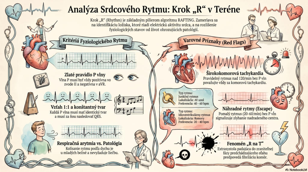
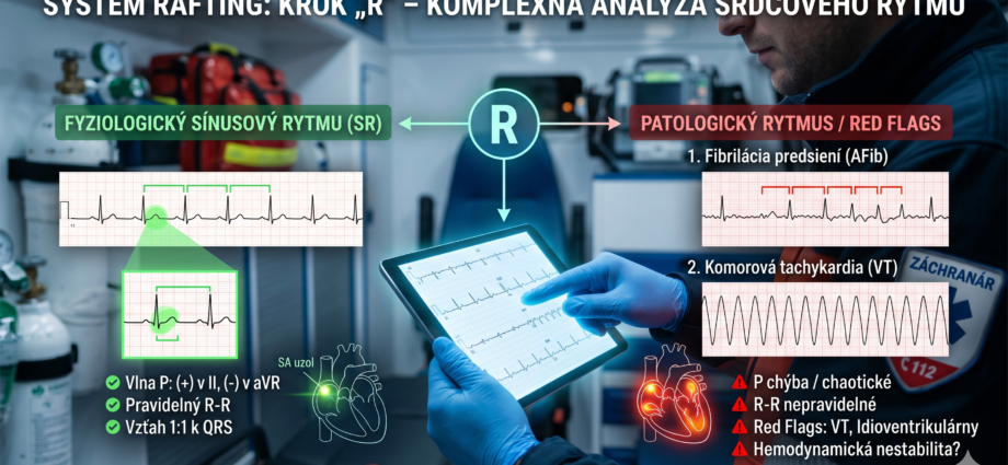
- Atrial depolarization vector: Since the impulse spreads from the SA node (located in the upper right atrium) downward and to the left toward the AV node, the resulting vector has an axis of approximately +60°. This gives an absolute rule: the P wave must be positive in lead II and negative in lead aVR.
- Constant morphology: P waves in a given lead must have an identical shape, confirming they originate from a single focus.
- 1:1 relationship: Every P wave must be followed by a QRS complex (with a constant PR interval). Paramedic context: If a patient shows P waves that are negative in II and positive in aVR, this is not sinus rhythm but an ectopic low-atrial or junctional rhythm with retrograde conduction.
2.2 Regularity vs. Irregularity: Looking for a Pattern
Measuring the distances between R waves (the R-R interval) determines rhythm regularity. Irregularity requires immediate differential diagnosis:
- Physiological (benign) irregularity: Respiratory sinus arrhythmia, where the rhythm speeds up on inspiration and slows on expiration. Common in children and young adults, requiring no treatment.
- Irregularly irregular rhythm: Absent P waves and chaotic R-R intervals are pathognomonic for atrial fibrillation (AFib). In older patients this is the most common arrhythmia, and it can lead to acute heart failure or cardiogenic shock if the ventricular response is inappropriately fast (Tachy-AFib).
- Regularly irregular rhythm: Occurs with 2nd-degree AV block (Wenckebach type) or with bigeminy/trigeminy (regular occurrence of extrasystoles).
2.3 Escape vs. Ectopic Rhythms
The paramedic must distinguish whether the heart is beating in a non-sinus rhythm because the SA node has failed (an escape mechanism), or because another focus is pathologically hyperactive (an ectopic mechanism).
- Junctional escape rhythm: If the SA node fails, the AV node takes over. It manifests as narrow QRS complexes at a rate of 40–60/min. P waves are either absent (hidden within the QRS) or retrograde (negative in II). Clinical significance: Often points to inferior-wall myocardial ischemia (RCA occlusion) or drug intoxication (digoxin, beta-blockers).
- Ventricular escape (idioventricular) rhythm: The last line of rescue. Rate 20–40/min, wide QRS complexes, absent P waves. Represents a state of imminent life threat and often precedes asystole.
3. RED FLAGS IN RHYTHM ANALYSIS
For prehospital practice, we define the following critical red flags at step “R” that must immediately shift the priority from assessment to treatment:
- Wide-complex tachycardia with no obvious P waves: Any regular wide-complex rhythm above 120/min in an adult patient must be treated in the field as ventricular tachycardia (VT) until proven otherwise. Searching for P waves must not delay preparation for cardioversion if the patient is hemodynamically unstable.
- Sudden-onset bradycardia with a narrow QRS and absent P waves: Points to dyssynchrony and SA node failure (often so-called Sick Sinus Syndrome). Risk of syncope and patient falls.
- A “flood” of ectopic beats (PVCs): The occurrence of polymorphic (varying-shape) ventricular extrasystoles, or the “R-on-T” phenomenon (an extrasystole falling within the vulnerable phase of the preceding beat), is a harbinger of malignant arrhythmia and ventricular fibrillation.
4. DISCUSSION
Precisely determining the underlying rhythm has a fundamental impact on the paramedic’s overall diagnostic conclusion. Misidentification leads to a cascade of errors. For example, if a paramedic fails to recognize a paced rhythm (the presence of “spikes” before the QRS and a wide complex) and applies criteria for sinus rhythm, they may falsely diagnose a bundle branch block or ST-segment elevation. Understanding the rhythm’s origin mechanism allows the paramedic not just to treat a symptom (e.g., giving atropine for bradycardia), but to anticipate how the condition will evolve and select adequately targeted pharmacotherapy (e.g., amiodarone vs. adenosine for tachycardias).
5. CONCLUSION
Step “R” – Rhythm is the entry gate to the algorithmic RAFTING system. It teaches paramedics not to perceive the ECG as merely a static image, but as a dynamic record of the heart’s electrophysiological hierarchy. Reliably distinguishing physiological sinus rhythm from pathological ectopy or escape rhythms directly shapes the treatment approach in terms of supporting hemodynamics and preventing sudden cardiac death in the prehospital phase.
6. REFERENCES
- DOBIÁŠ, V. et al. (2021). Prehospital Emergency Medicine. 3rd edition. Martin: Osveta.
- HAMPTON, J. R. (2019). The ECG Made Easy. 9th edition. Elsevier.
- TÁBORSKÝ, M. et al. (2021). Clinical Cardiology. Mladá fronta. (Supplementary literature on deep electrophysiological mechanisms.)
- Guidelines of the European Resuscitation Council (ERC) for the management of peri-arrest arrhythmias.
- Slovak Ministry of Health guidance on the provision of prehospital emergency medical care.