Lung Auscultation —
technique, examination points, phenomena
The stethoscope is the most-used and simultaneously the most misused instrument in a paramedic’s bag. The difference between “I heard something” and precise localization and classification of a breath sound phenomenon determines whether the auscultatory finding is clinically usable.
Building on VYS-01 (IPPA order) and VYS-02 (field limits), this installment covers lung auscultation in detail — correct stethoscope diaphragm and bell placement technique, the systematics of examination points on the chest, distinguishing physiological from pathological breath sound phenomena, and the specific limits imposed by ambulance noise while driving.
The stethoscope — diaphragm vs. bell
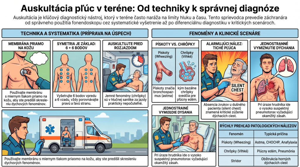
Diaphragm (the flat side) — transmits high-frequency sounds; used for breath sound phenomena and most heart sounds. Applied with gentle pressure.
Bell (the cup-shaped side) — transmits low-frequency sounds; used mainly for heart auscultation (see VYS-06). Applied with light touch, without pressure — firm pressure turns the bell into a diaphragm.
For lungs, the diaphragm is used as standard — breath sound phenomena predominantly have a higher frequency than most pathological heart sounds.
Systematics of examination points
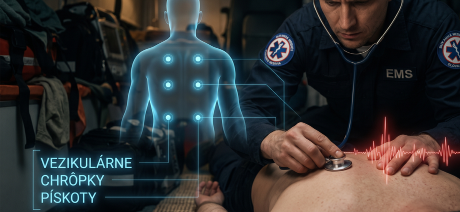
Auscultating the lungs without systematics leads to only easily accessible areas being examined (the upper anterior chest) while the lung bases — where crackles from congestion are heard most often — or the axillary areas important for middle-lobe pneumonia are skipped.
Auscultating through thin clothing distorts high-frequency sounds and can create artifacts (fabric rustling mimicking crackles). Whenever the situation and time allow, the stethoscope is applied directly to the skin. In cold weather or a mass casualty incident where there’s no time to expose the chest, this compromise must be factored into interpreting the finding.
Breath sound phenomena — classification
| Phenomenon | Sound characteristics | Typical cause |
|---|---|---|
| Vesicular breathing | Physiological finding — a soft, “rustling” sound, longer inspiration than expiration | Normal finding |
| Crackles (wet phenomena) | Crackling, discontinuous sounds, predominantly in inspiration | Congestion, pulmonary edema, pneumonia |
| Rhonchi (dry phenomena) | Low-frequency, continuous, “rattling” | Secretions in larger airways, bronchitis |
| Wheezes | High-frequency, whistling, predominantly in expiration | Bronchospasm — asthma, COPD, anaphylaxis |
| Stridor | Loud, high-frequency, audible even without a stethoscope, predominantly in inspiration | Upper airway obstruction — an urgent finding |
| Pleural friction rub | “Creaking,” like walking on snow, synchronous with breathing | Pleuritis, pleurisy |
| Diminished to absent breath sounds | Reduced intensity or complete absence of breath sounds | Pneumothorax, pleural effusion, atelectasis, deep single-bronchus intubation |
The limits of auscultation in a noisy environment
As already noted in VYS-02, auscultation is the most vulnerable of all four pillars of physical examination to ambient noise. While an ambulance is driving in ordinary traffic, reliable auscultation of subtle breath sound phenomena (particularly lower-intensity crackles) is practically impossible.
Auscultation before departure — if the patient’s condition allows, a complete auscultation is performed on scene or with the vehicle stopped before departure, not while driving.
Targeted auscultation during transport — while driving, only a gross, loud finding can realistically be reliably assessed (marked wheezing, absent breathing on one side), not subtle differences.
Electronic stethoscopes — part of some crews’ equipment, they amplify the signal and partially suppress ambient noise; their use is worth considering particularly for noisier calls.
Visual and palpatory substitutes — when reliable auscultation isn’t possible, the finding is supplemented by inspection (symmetry of chest movement, use of accessory muscles) and palpation (fremitus), which are more resistant to noise.
Three scenarios from practice
A young patient with sudden dyspnea, wheezing audible even without a stethoscope, prolonged expiration.
A 78-year-old female patient with worsening dyspnea, orthopnea, lower-extremity edema.
A patient after a stab injury to the left hemithorax, growing dyspnea.
What the next installment covers
Installment VYS-05 continues with chest examination as a whole — inspection, palpation, and percussion of the chest wall, recognizing rib fractures, subcutaneous emphysema, and paradoxical breathing in flail chest.
- VYS-01 — Inspection, Palpation, Percussion, Auscultation: The Four Pillars of Physical Examination
- VYS-02 — Physical Examination in the Field vs. the Hospital
- VYS-03 — Head and Neck Examination
- VYS-04 — Lung Auscultation (this article)
- VYS-05 — Chest Examination
- VYS-06 — Heart Auscultation
- VYS-07 — Abdominal Examination
- VYS-08 — Peritoneal Signs and Acute Abdomen
- VYS-09 — Examination of the Limbs and Peripheral Circulation
- VYS-10 — Neurological Examination in the Field
- VYS-11 — The Skin as a Diagnostic Tool
- VYS-12 — Examining the Pediatric Patient
- VYS-13 — Examining the Geriatric Patient
- VYS-14 — From Examination to Decision
- Bickley LS, Szilagyi PG, Hoffman RM. Bates’ Guide to Physical Examination and History Taking. 13th ed. Wolters Kluwer, 2021 — chapter on chest and lung examination.
- Sarkar M, Madabhavi I, Niranjan N, Dogra M. Auscultation of the respiratory system. Annals of Thoracic Medicine. 2015;10(3):158–168.
- Bohadana A, Izbicki G, Kraman SS. Fundamentals of lung auscultation. New England Journal of Medicine. 2014;370:744–751.
- National Association of EMTs (NAEMT). PHTLS — Prehospital Trauma Life Support. 10th ed., 2023.
- Global Initiative for Asthma (GINA). Global Strategy for Asthma Management and Prevention, 2023 — assessing severity by auscultatory finding.