
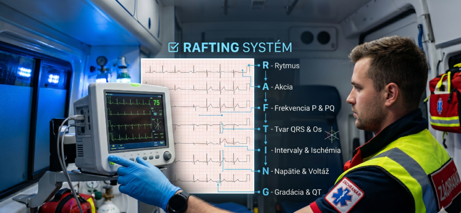
The RAFTING System, Step “T”: QRS Morphology and Electrical Axis
QRS width decides whether you are looking at a conduction disorder, a ventricular rhythm, or a tracing that cannot be read reliably without an older ECG for comparison.
After rhythm, rate, and the analysis of atrial activity with AV conduction, one question remains in the core RAFT sequence: how do the ventricles depolarise? Step “T” (Type of QRS & Axis) assesses the width and shape of the QRS complex and the electrical axis of the heart. This is the step where the field decision is most often made: is the tracing safe for transport, or do you need to prepare the defibrillator and call a physician?
Where step T fits in the RAFTING system
The introductory part of the series defines RAFTING as a sequential checklist that should lead a paramedic through a tracing in the same order, even under pressure. Steps R, A and F cover rhythm, rate, and the atria with AV conduction. Step T turns to the ventricles: how long their activation takes, what it looks like, and in which direction it spreads. The introductory part also lists the later steps I, N and G (repolarisation, voltage, general impression and QT); this article does not cover them.
1. QRS width (narrow or wide complex), 2. QRS shape in leads V1 and V6 (right or left bundle branch block, nonspecific widening) and 3. the electrical axis in the frontal plane. The detailed method for determining the axis is in the separate part Determining the Electrical Axis of the Heart; here only its place in the system is described.
Illustrative scenario. The patient is sitting, conscious, BP 148/86, SpO₂ 96 %, heart rate 84/min. He describes pressure behind the sternum since the morning. The 12-lead ECG shows sinus rhythm, QRS 148 ms, a deep S in V1, a broad monophasic R without a q wave in V6, and an axis of about −10°. No older tracing is available.
Decision: the tracing is read as left bundle branch block with no way to tell whether it is new. The crew does not treat the block as a benign finding, because the clinical picture points to an acute coronary syndrome. They send the tracing to the cardiac centre and continue monitoring.
1. QRS width: the first measurement
At the standard paper speed of 25 mm/s, one small square equals 40 ms, so three squares equal 120 ms. Measure from the start of the Q or R wave to the end of the S wave in the lead where the complex is best defined. The value printed by the device is useful but does not replace your own measurement: automatic analysis is often least accurate precisely with wide complexes.
| QRS width (adult) | Term | What it means for the next step |
|---|---|---|
| up to 110 ms | Narrow complex | The ventricles are activated through the conduction system; continue with the axis and the remaining steps. |
| 110–119 ms | Borderline widening | Incomplete bundle branch block or nonspecific widening; judge the shape in V1 and V6. |
| 120 ms or more | Wide complex | Bundle branch block, ventricular origin, pre-excitation, pacing, or a metabolic or toxic cause; always read it in context. |
The 120 ms threshold for complete bundle branch block in adults comes from the AHA/ACCF/HRS statement on ECG standardisation, part III [1]. The 110–119 ms range is associated with incomplete block in the same document.
2. QRS shape: right and left bundle branch block
With a wide QRS, look at two leads: V1 and V6. Their shape usually tells you which bundle branch conducts late.
| Feature | Right bundle branch block (RBBB) | Left bundle branch block (LBBB) |
|---|---|---|
| V1 | rSR′ or rsR′ (“rabbit ears”) | deep S or QS |
| I and V6 | wide, slurred S | wide, notched or flat-topped R, no Q |
| QRS width | 120 ms or more | 120 ms or more |
| ST-T assessment | secondary changes in the right precordial leads | ST-T discordant to the QRS; see the infarction section |
Descriptions and criteria follow the AHA/ACCF/HRS statement [1]. Isolated right bundle branch block is often a finding without clinical significance in older patients; left bundle branch block is more often associated with structural heart disease. A paramedic does not interpret these associations as a diagnosis. The task is to recognise the block, record it, and alert the physician that the finding may mask other changes.
A similar tracing can come from a paced rhythm (pacing spike before the complex, shape often resembling LBBB), a ventricular rhythm or ectopic beats, pre-excitation with a delta wave and a short PR interval, as well as metabolic and toxic causes. The ERC lists hyperkalaemia among the reversible causes of cardiac arrest and deteriorating arrhythmias [7]. With a wide QRS and bradycardia, or in an unstable patient, consider these causes too and contact a physician immediately.
3. Electrical axis: a brief placement
In step T the axis is determined with the quick quadrant method from leads I and aVF. This is not a precise measurement in degrees; it only establishes whether the axis falls into a pathological quadrant.
- Look at leads I and aVF. A lead is positive when the complex points mainly upward.
- I and aVF both positive: the axis is normal.
- I positive, aVF negative: deviation to the left. If lead II is still positive, the axis is up to −30° and still normal; if II is negative, this is left axis deviation.
- I negative, aVF positive: deviation to the right.
- I and aVF both negative: extreme axis, which is very rare.
A negative P wave, QRS and T wave in lead I together with a positive aVR most often means the right and left arm electrodes were swapped, not true axis deviation or dextrocardia. Check the limb electrode placement before drawing any conclusion about the axis, and repeat the recording if in doubt.
Left axis deviation with a narrow QRS may correspond to left anterior fascicular block (qR in aVL, QRS under 120 ms) [1]. In a patient with sudden dyspnoea, right axis deviation raises the suspicion of right ventricular strain; the finding is nonspecific and is always judged together with the clinical picture, never on its own.
4. A wide QRS during tachycardia
Distinguishing ventricular tachycardia from supraventricular tachycardia with aberrant conduction is difficult even for physicians. The 2019 ESC guidelines on supraventricular tachycardia advise managing a wide-complex tachycardia of unclear origin as ventricular tachycardia [6]. For a paramedic, the algorithm must not replace the decision about the patient: haemodynamic instability and adverse features determine the course of action regardless of the result of the shape analysis.
In 1991 Brugada and colleagues described a four-step algorithm for regular wide-complex tachycardia; in the original study of 554 tachycardias it reached a sensitivity of 98.7 % and a specificity of 96.5 % [2]. A later algorithm using lead aVR alone achieved higher accuracy than the Brugada algorithm according to its authors [3]. Both are aids to recognition, not to prescribing treatment.
An irregular wide complex with a very fast rate and varying QRS shape may represent atrial fibrillation conducted over an accessory pathway in pre-excitation. Giving drugs that slow AV nodal conduction is a risky choice in this situation; the 2019 ESC guidelines do not recommend them for pre-excited atrial fibrillation [6]. The choice of drug and of electrical cardioversion is made by the physician according to the patient’s condition; see step “F” for the related discussion of a short PR interval and the delta wave.
5. Left bundle branch block and suspected infarction
Left bundle branch block alters the course of the ST segment and can mask changes of acute myocardial infarction. In 1996 Sgarbossa and colleagues, using data from the GUSTO-1 trial, described three independent criteria: ST elevation of at least 1 mm concordant with the QRS, ST depression of at least 1 mm in V1–V3, and ST elevation of at least 5 mm discordant to the QRS [4]. In 2012 Smith and colleagues proposed replacing the absolute 5 mm threshold with the ratio of ST elevation to the depth of the S wave (less than −0.25); in their series of 33 patients with an occluded coronary artery, the modified rule had a sensitivity of 91 % and a specificity of 90 % [5]. This is a small retrospective series and the results should be interpreted with caution.
With known or new left bundle branch block and a clinical suspicion of acute coronary syndrome, do not look for ST elevation by the usual STEMI criteria. Record any concordant changes, send the tracing to the cardiac centre via the CDS App, and leave the decision about intervention to the cardiologist. Comparison with an older tracing is also important, if the patient or family has one.
How to document step T
“ECG: block, patient stable.”
“12-lead ECG at 14:32: sinus rhythm 84/min, QRS 148 ms, morphology consistent with left bundle branch block, axis about −10°. No older tracing available. Tracing sent to the cardiac centre via the CDS App at 14:35.”
The scope of practice of a paramedic, including recording and assessing the ECG while providing emergency care, is regulated by Decree of the Ministry of Health of the Slovak Republic No. 321/2005 Coll., as amended. The paramedic uses the assessment to decide on the course of action and to communicate with the physician. The diagnostic conclusion and specific antiarrhythmic treatment belong to the physician; verify the wording of the regulation in its current version on slov-lex.sk.
Decree of the Ministry of Health of the Slovak Republic No. 321/2005 Coll.
Practical implications for the paramedic
1. Always measure QRS width yourself; three small squares at 25 mm/s are 120 ms.
2. With a wide QRS, look at V1 and V6 and tell right bundle branch block, left bundle branch block, and nonspecific widening apart.
3. A wide QRS is not yet a block: think of pacing, ventricular origin, pre-excitation, hyperkalaemia and toxic causes.
4. Treat a regular wide-complex tachycardia as ventricular; the unstable patient sets the course of action, not the shape analysis.
5. With left bundle branch block and suspected acute coronary syndrome, send the tracing to the cardiac centre and compare it with an older ECG.
6. Do not trust the axis with a negative lead I and a positive aVR until you have ruled out electrode reversal.
- The biological basis of the ECG
- Determining the electrical axis of the heart
- The RAFTING system: algorithmic ECG interpretation
- Step “R” — heart rhythm analysis
- Step “A” — heart rate
- Step “F” — atrial activity and AV conduction
- Step “T” — QRS morphology and electrical axis
- Steps “I”, “N” and “G” — ischaemia, voltage, QT and general impression
- Surawicz B, Childers R, Deal BJ, Gettes LS, Bailey JJ, Gorgels A, et al. AHA/ACCF/HRS recommendations for the standardization and interpretation of the electrocardiogram: part III: intraventricular conduction disturbances. Circulation. 2009;119(10):e235–e240. doi:10.1161/CIRCULATIONAHA.108.191095
- Brugada P, Brugada J, Mont L, Smeets J, Andries EW. A new approach to the differential diagnosis of a regular tachycardia with a wide QRS complex. Circulation. 1991;83(5):1649–1659. doi:10.1161/01.cir.83.5.1649
- Vereckei A, Duray G, Szénási G, Altemose GT, Miller JM. New algorithm using only lead aVR for differential diagnosis of wide QRS complex tachycardia. Heart Rhythm. 2008;5(1):89–98. doi:10.1016/j.hrthm.2007.09.020
- Sgarbossa EB, Pinski SL, Barbagelata A, Underwood DA, Gates KB, Topol EJ, et al. Electrocardiographic diagnosis of evolving acute myocardial infarction in the presence of left bundle-branch block. N Engl J Med. 1996;334(8):481–487. doi:10.1056/NEJM199602223340801
- Smith SW, Dodd KW, Henry TD, Dvorak DM, Pearce LA. Diagnosis of ST-elevation myocardial infarction in the presence of left bundle branch block with the ST-elevation to S-wave ratio in a modified Sgarbossa rule. Ann Emerg Med. 2012;60(6):766–776. doi:10.1016/j.annemergmed.2012.07.119
- Brugada J, Katritsis DG, Arbelo E, Arribas F, Bax JJ, Blomström-Lundqvist C, et al. 2019 ESC Guidelines for the management of patients with supraventricular tachycardia. Eur Heart J. 2020;41(5):655–720. doi:10.1093/eurheartj/ehz467
- Soar J, Böttiger BW, Carli P, Couper K, Deakin CD, Djärv T, et al. European Resuscitation Council Guidelines 2021: Adult advanced life support. Resuscitation. 2021;161:115–151. doi:10.1016/j.resuscitation.2021.02.010
- Decree of the Ministry of Health of the Slovak Republic No. 321/2005 Coll. on the scope of practice of certain health professions, as amended.