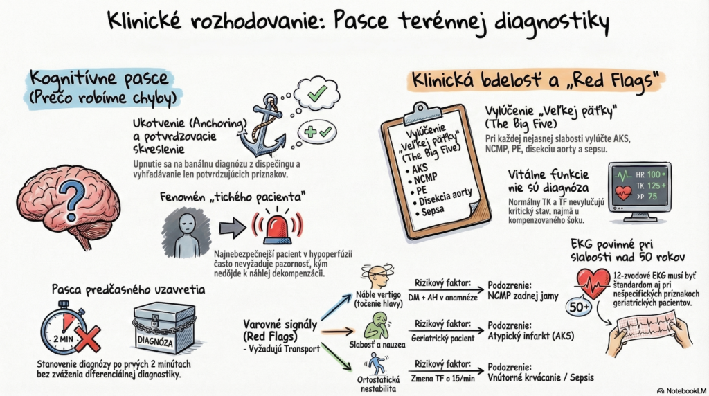
In Slovak prehospital emergency care, clinical decision-making is a process that unfolds under time pressure, in a non-standard environment, and often with limited information. This article examines the critical points where a discrepancy arises between the protocol-driven procedure and the paramedic’s subjective gut feeling.
1. INTRODUCTION – A REALISTIC SCENARIO
Dispatch: Basic ambulance, 09:15, male, 64 years old, “weakness, dizziness.” On scene: The patient is sitting in an armchair, alert, oriented, calmly conversational. He reports that on getting up this morning he felt unsteady while walking and had mild nausea. He now feels better and only wants his “blood pressure checked.” Objective findings: Skin warm, dry, no cyanosis. Breathing unlabored. BP: 155/95 mmHg, HR: 88/min (regular), SpO2: 96% (room air), GCS: 15, Temp: 36.8°C. History: Stage II hypertension, type 2 diabetes on oral hypoglycemics. Crew’s assessment: “The patient doesn’t look sick.” Paperwork for leaving him on scene begins.
2. CLINICAL ANALYSIS OF THE SITUATION (an ABCDE approach)
Although vital signs show no sign of shock or respiratory failure, a static set of readings is only a “snapshot,” not a “film” of the patient’s condition.
- A/B (Airway/Breathing): No obvious pathology, but a quiet patient with nausea can be masking a silent aspiration or pulmonary embolism (PE).
- C (Circulation): BP and HR are within normal limits, but for a diabetic patient with a history of hypertension, 155/95 mmHg could represent relative hypotension compared to his usual BP of 180/100 mmHg.
- D (Disability): The key point. Weakness and nausea in an elderly diabetic patient are highly non-specific, but alarming symptoms (an anginal equivalent, posterior fossa stroke, aortic dissection).
- E (Exposure): The abdominal exam (pulsatile mass?) or lower-limb exam (asymmetry?) is often neglected in a “dizziness” call.
Red flags in this scenario:
- Sudden-onset unsteadiness while walking (vertigo).
- A high-risk profile (diabetes + hypertension).
- A mismatch between subjective improvement and the mechanism behind the symptoms.
3. THE MOST COMMON ERRORS IN PRACTICE
In the field, Slovak EMS crews battle cognitive phenomena that distort objectivity:
- Anchoring: The paramedic fixates on the dispatch information (“high blood pressure”) and ignores new symptoms that don’t fit a hypertension diagnosis.
- Confirmation bias: Looking only for signs that confirm a benign diagnosis, in order to avoid an “unnecessary” transport.
- Premature closure: A diagnosis is settled within the first 2 minutes without any differential-diagnostic reasoning.
- The quiet patient: The most dangerous type. While a loud, distressed patient demands attention, a quiet patient in hypoperfusion or with internal hemorrhage slips under the radar until sudden decompensation.
4. DECISION POINTS (CRITICAL THINKING)
When must the original plan change?
- Trend of the condition: If vital signs change during assessment (e.g., HR rises by 15/min with positional change), this indicates orthostatic instability — a clear indication for transport.
- “Gut feeling” vs. EBM: An experienced paramedic’s intuition is a lightning-fast, subconscious pattern-recognition process. If you feel “something’s off” despite normal vitals, transport.
- Diagnostic uncertainty: If in the field you cannot rule out a life-threatening condition (e.g., MI in a diabetic with epigastric pain), the diagnosis reads: “Suspected acute condition — requires hospital-based diagnostics.”
5. A PRACTICAL APPROACH FOR BASIC AMBULANCE CREWS
Apply this algorithm to every “trivial” call:
- Complete monitoring: 12-lead ECG (mandatory for any weakness in a patient over 50!), blood glucose, SpO2, blood pressure on both arms.
- SAMPLE history: Focus on last meal/last output and any changes in medication (anticoagulants!).
- Stability test: If the patient reports weakness, don’t just let them sit. Try positional change (if safe) and watch the circulatory response.
- Differential diagnosis (ruling out “the big five”):
- Acute coronary syndrome (ACS).
- Stroke.
- Pulmonary embolism (PE).
- Aortic dissection.
- Sepsis / internal hemorrhage.
6. PHARMACOLOGICAL AND INTERVENTIONAL CONSIDERATIONS
For unclear presentations, within basic-crew scope:
- Crystalloids: For relative hypotension (as long as there’s no sign of pulmonary edema) — a 250–500 ml bolus to test the circulatory response.
- Antiplatelet therapy (aspirin): For any suspicion of ACS, give 250–500 mg (chewed PO or IV per local protocol).
- Antiemetics (metoclopramide): Caution — they can mask the symptoms of an intra-abdominal catastrophe or neurological deficit. Give only with a clear etiology.
7. TAKE-HOME MESSAGE
- Vital signs are not a diagnosis: Normal BP and HR do not rule out an ongoing critical condition (e.g., compensated shock).
- Suspicion is enough: The EMS crew’s job is not to establish a definitive diagnosis, but to triage and stabilize safely.
- Documentation: If you leave a patient at home, the record must clearly state why the condition was assessed as non-serious and what specific instructions the patient was given.
- Atypical presentation: Elderly and diabetic patients may not show textbook symptoms (e.g., a “silent” heart attack).
8. BONUS – “HOW THIS COULD GO WRONG”
Alternative ending: The crew assessed the condition as a “hypertensive reaction to stress,” gave 25 mg sublingual captopril, and left the patient at home. Two hours later, family calls again — the patient is unconscious.
Reality: It was a basilar artery thrombosis (a posterior fossa stroke). The weakness and nausea were prodromal symptoms. The result is irreversible brainstem damage.
Consequence: A forensic follow-up, a failure in the differential-diagnosis process, and disregard for the patient’s risk factors.
Sources:
- Slovak Ministry of Health guidance on the provision of emergency healthcare.
- ERC Guidelines 2021 / ALS protocols.
- Clinical protocols for Slovak EMS (EMS Operations Center).
CHECKLIST: Differential diagnosis of non-specific weakness (basic crew)
This list serves to rule out critical conditions before deciding to leave a patient on scene or transport to a less specialized facility.
1. History review (risk stratification)
- [ ] Age over 65? (Increases risk of atypical ACS/stroke presentation.)
- [ ] Diabetes mellitus? (Risk of silent myocardial ischemia and serious metabolic imbalance.)
- [ ] Anticoagulant therapy? (Warfarin, NOACs — risk of intracranial or occult hemorrhage even after minor trauma.)
- [ ] Oncology history? (Risk of pulmonary embolism, metabolic derangement.)
- [ ] Sudden onset? (Onset over seconds/minutes suggests a vascular or mechanical cause.)
2. Objective exam (the “big five” filter)
- [ ] ECG (12-lead): Are ST elevations/depressions, a bundle branch block (LBBB), or a new arrhythmia present?
- [ ] Neurological screening (FAST/G-FAST): Is there facial asymmetry, limb weakness, speech disturbance, or gaze deviation?
- [ ] Orthostatic test: If BP drops more than 20 mmHg or HR rises more than 20/min on standing, this points to clear dehydration or occult shock.
- [ ] Abdominal palpation: Is there a pulsatile mass (AAA) or a palpable hernia?
- [ ] Lower limbs: Is there unilateral swelling or calf pain (suspicion of venous thromboembolism)?
3. Lab/technical mini-panel
- [ ] Blood glucose: Rule out hypo/hyperglycemia (a normal value doesn’t mean the patient is fine, but it does rule out one cause).
- [ ] SpO2 on both arms: A significant difference can indicate aortic dissection.
- [ ] Body temperature: Rule out early sepsis in an elderly patient (confusion and weakness may be present without fever).
4. Cognitive check (anti-bias review)
- [ ] “Doesn’t look sick”: Did I ask the patient how they feel compared to their normal day?
- [ ] “Quiet patient”: Are they calm because nothing is wrong, or because they’re apathetic/in shock?
- [ ] “Routine”: Did I follow ABCDE, or did I settle on a diagnosis already “at the door”?
Decision algorithm (triage)
- Red zone (immediate transport):
- An abnormal ECG (STE, NSTE, arrhythmia with low cardiac output).
- Positive FAST or a new neurological deficit.
- Uncorrected hypotension or tachycardia.
- Severe pain (chest, abdomen, back).
- Yellow zone (transport for further workup):
- Unclear etiology of weakness in a high-risk patient (diabetic, elderly).
- Inability to walk unassisted (if previously able).
- Persistent nausea and vomiting of unclear origin.
- Green zone (leave on scene — extreme caution):
- A clear etiology (e.g., confirmed gastroenteritis in the family).
- Stable vital signs, negative 12-lead, negative neurological findings.
- The patient’s ability to understand instructions, and a third party present.
Take-home message for practice:
If you decide to leave a patient on scene, your record must include the statement: “The patient was instructed to call EMS again if their condition worsens, if chest pain develops, or if a neurological deficit appears.” Under Slovak law, this documented instruction is your primary protection.