ABSTRACT
The third step of the RAFTING diagnostic algorithm, represented by the letter “F” (Frequency of P & PR interval / Atrial activity and conduction), shifts the examiner’s attention from the prominent ventricular complex to the subtler, yet clinically equally important, manifestations of atrial depolarization. This article discusses in detail the significance of P-wave morphology analysis for indirectly diagnosing atrial pressure or volume overload. It then defines the pathophysiology of atrioventricular (AV) conduction, measured via the PQ (PR) interval. Systematic evaluation of this interval allows the paramedic to identify impending higher-degree AV blocks early and to anticipate sudden hemodynamic destabilization of the patient in prehospital care.
1. INTRODUCTION
In the dynamic and stressful environment of prehospital emergency medical care, so-called “tunnel vision” is common, where the paramedic analyzes only the dominant deflections of the ECG trace — the QRS complex and the ST segment. The P wave and its following isoelectric segment, however, conceal vital information about the heart’s structural state and the integrity of its electrical wiring. Step “F” in the RAFTING system teaches paramedics that the signal delay in the AV node isn’t a fault, but a necessary physiological mechanism that ensures adequate time for the ventricles to fill mechanically. A disturbance of this mechanism, however, turns the ECG into a ticking time bomb.
2. CORE: MORPHOLOGY AND CONDUCTION
2.1 P-Wave Morphology: A Window into the Atria
The P wave represents depolarization of the right and then the left atrium. Under normal circumstances it’s smooth and gradual. In prehospital care we look for two specific pathologies that indicate atrial overload:
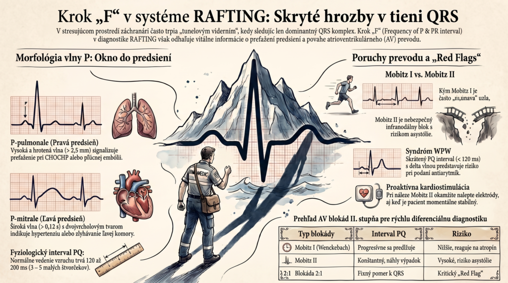
- P-pulmonale (right atrial overload): The P wave is abnormally tall and peaked (more than 2.5 mm, i.e., more than half a large square in height, in lead II). This finding indicates elevated pressure in the pulmonary circulation, against which the right heart must work. In emergency medicine this is a typical picture in chronic obstructive pulmonary disease (COPD), severe asthma, or acutely in massive pulmonary embolism (so-called cor pulmonale).
- P-mitrale (left atrial overload): The P wave is wide (over 0.12 s) and often has a double-humped, “camel-back” shape (notch wider than 1 small square). It reflects delayed depolarization of an enlarged left atrium. In the field, this is seen in patients with long-standing untreated arterial hypertension, valvular disease (mitral stenosis), or left ventricular failure.
2.2 The PQ (PR) Interval and Its Physiology
The PQ interval is measured from the start of the P wave to the start of the QRS complex. It encompasses the time from impulse origin in the SA node, through its passage across the atria, and particularly its slowing in the AV node.
- Physiological range: 120 to 200 ms (3 to 5 small squares at 25 mm/s paper speed).
2.3 Conduction Pathology: Atrioventricular Blocks
The paramedic must categorize conduction disorders by severity and location of the block:
- Prolonged PQ interval (> 200 ms) – 1st-degree AV block: The impulse always reaches the ventricles, but it takes longer. This is a constant prolongation. Often a benign finding (athletes, medication effect); by itself it requires no treatment, but signals altered conduction system function.
- Progressively lengthening PQ with a dropped beat – 2nd-degree AV block, Mobitz I (Wenckebach): The PQ interval progressively lengthens with each beat, until one impulse fails to pass through the AV node at all (on the ECG we see a P wave not followed by a QRS). The block is usually within the AV node itself, caused by cell fatigue, and often responds well to atropine. Typical of acute inferior-wall infarction.
- Constant PQ with a sudden dropped beat – 2nd-degree AV block, Mobitz II: A treacherous and dangerous type. The PQ interval is constant (normal or prolonged), but the QRS complex suddenly drops out entirely. The block is located infranodally (in the bundle of His or the bundle branches). It carries a high risk of sudden progression to complete block (asystole).
- Shortened PQ interval (< 120 ms): The signal bypasses the slowing AV node via a pathological accessory pathway. If a delta wave is also present (a slurred upstroke of the QRS), this is Wolff-Parkinson-White (WPW) syndrome.
3. RED FLAGS IN PREHOSPITAL PRACTICE
Step “F” generates the following warning signals for the paramedic:
- A finding of Mobitz II AV block or 2:1 block: This is an absolute red flag. Even if the patient is currently hemodynamically stable, the paramedic must immediately apply multifunction pacing pads and be ready for transcutaneous pacing. Giving rate-slowing drugs (amiodarone, beta-blockers) is strictly contraindicated.
- New-onset P-pulmonale in a dyspneic patient: If the patient is suddenly short of breath, hypoxic, and a tall peaked P wave appears in lead II, differential diagnosis must immediately focus on acute pulmonary embolism, particularly if tachycardia is also present.
- A shortened PQ interval in a tachycardic patient: A patient with palpitations and wide-complex tachycardia who has a history (or an older ECG) showing a shortened PQ interval may have atrial fibrillation conducting via a WPW pathway. Giving standard drugs (such as adenosine or verapamil) can block the AV node, conduct all impulses via the accessory pathway, and trigger fatal ventricular fibrillation. Electrical cardioversion is preferred.
4. DISCUSSION
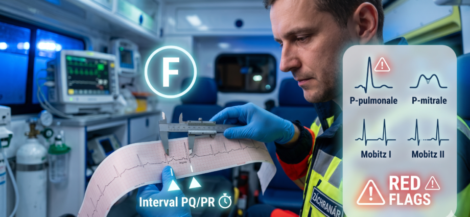
In paramedic practice, assessing the PQ interval is often pushed aside because of its relative visual inconspicuousness. A dropped QRS complex during a Wenckebach period is frequently misinterpreted as a sinus pause or a blocked atrial extrasystole. This mistake can lead to incorrect antiarrhythmic administration. Consistently applying step “F” of the RAFTING system forces the examiner to pick up an ECG ruler (or calipers) and precisely measure distances. Only this way can the hidden threat of infranodal blocks be uncovered — blocks that could cause sudden loss of consciousness (Adams-Stokes syndrome) at any time during transport.
5. CONCLUSION
Analyzing atrial activity and the PQ interval is a critical tool in prehospital care for assessing the risk of sudden conduction-system failure. A paramedic who has mastered step “F” doesn’t wait for cardiogenic shock or asystole to develop. They can recognize warning changes (Mobitz II, extreme PQ prolongation) at a stage when the patient is still conscious, and proactively prepare pacing equipment, dramatically increasing the patient’s chances of survival without neurological deficit.
6. REFERENCES
- DOBIÁŠ, V. et al. (2021). Prehospital Emergency Medicine. 3rd edition. Martin: Osveta.
- European Resuscitation Council (ERC). (2021). Guidelines for Resuscitation: Peri-arrest arrhythmias.
- HAMPTON, J. R. (2019). The ECG Made Easy. 9th edition. Elsevier.
- TÁBORSKÝ, M. et al. (2021). Clinical Cardiology. Mladá fronta.
- ŠANDRIKOVÁ, V. (2018). Electrocardiography for Non-Physician Healthcare Professions.