Capnography in the spontaneously
breathing patient
Opioid intoxication, status epilepticus, DKA — three scenarios where a nasal capnography cannula will change your management before the pulse oximeter reacts.
The previous five installments focused mainly on intubated patients and CPR. This final installment of the series closes the loop — we return to the spontaneously breathing patient and show where a nasal capnography cannula provides information that a pulse oximeter simply can’t. Three clinical scenarios every paramedic encounters on every shift.
Why a nasal cannula — a technical note
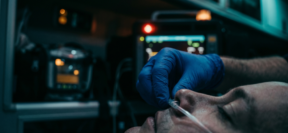
A sidestream capnograph allows EtCO₂ measurement via a nasal cannula in any spontaneously breathing patient — without intubation, without an invasive procedure. The cannula is inserted just like a standard oxygen cannula, with the difference that one port draws exhaled air and carries it through tubing to the measuring chamber.
An important technical limitation: EtCO₂ values from a nasal sample are less accurate than from intubation — exhaled air mixes with ambient air, and real values can be 2–5 mmHg lower than actual PaCO₂. Nevertheless, the nasal cannula has a fundamental clinical advantage: it provides a continuous trend — and it’s the trend, not the absolute number, that’s decisive in prehospital practice.
Every patient with altered consciousness, respiratory distress, or a risky pharmacological profile should have EtCO₂ monitoring from first contact. In practice, this means: just as routinely as SpO₂.
Corpuls 3, Zoll X Series, Lifepak 15 — all the monitors common in Slovak EMS have sidestream EtCO₂ as a standard feature. Nasal cannulas should be part of every crew’s kit, just like the BVM.
Scenario 1 — Opioid intoxication
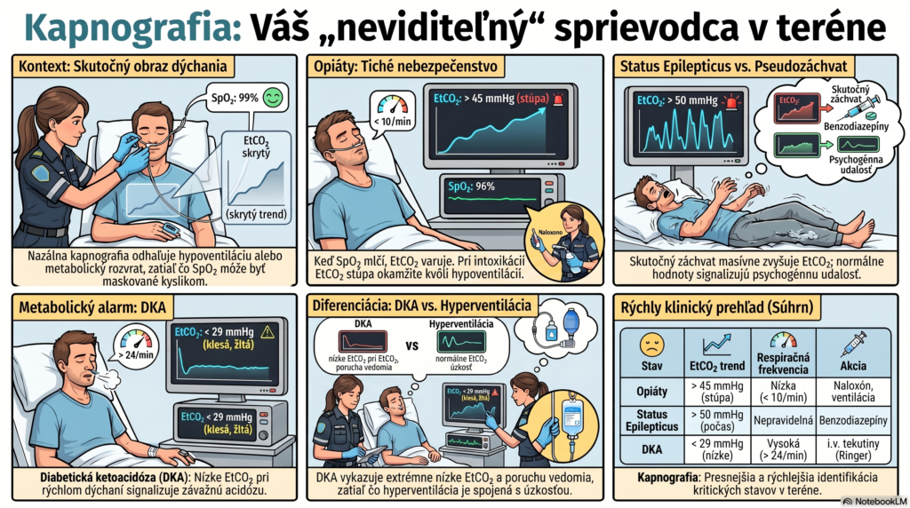
Opioid intoxication is a classic example of a situation where SpO₂ lies and EtCO₂ tells the truth. The mechanism is physiologically simple: opioids suppress the respiratory center in the medulla — the patient hypoventilates, CO₂ accumulates. EtCO₂ rises immediately. SpO₂ stays normal or only mildly reduced — particularly if the patient is on supplemental oxygen — until a critical hypoventilation crisis occurs.
The nasal capnography cannula shows: EtCO₂ 52 mmHg, RR 8/min, spread-out waveforms. SpO₂ still 96%. This is a classic hypoventilation trap — oxygenation masked by supplemental O₂, but ventilation is failing.
The paramedic reduces the O₂ flow, verbally stimulates the patient, prepares the BVM. Three minutes later, EtCO₂ = 58 mmHg, RR 6/min → titrated naloxone 0.2 mg IV is given. EtCO₂ drops to 44 mmHg within 90 seconds. Without capnography, the hypoventilation would have gone undetected.
Capnographic profile of opioid intoxication
| Parameter | Finding | Action |
|---|---|---|
| EtCO₂ | > 45 mmHg, progressively rising | Stimulate, reduce O₂, BVM if RR < 8/min |
| RR | < 10/min, slow spread-out waveforms | Naloxone 0.2–0.4 mg IV, titrated per protocol |
| Waveform shape | Square, but spread out — slow rate | Monitor the trend after naloxone — rebound risk |
| SpO₂ | Normal or mildly reduced — unreliable with O₂ | Don’t use as the primary indicator in opioid cases |
Scenario 2 — Status epilepticus
Status epilepticus (SE) brings capnography to the forefront at two moments: during the seizure itself and after giving benzodiazepines. Both deserve attention.
During the seizure
A generalized tonic-clonic seizure causes a massive increase in muscular metabolism — CO₂ production rises dramatically. EtCO₂ during an active seizure can reach 60–80 mmHg. This is a physiological response, not a pathological finding — but it signals an extreme metabolic burden on the brain.
Practical consequence: if EtCO₂ during a presumed seizure remains normal (35–45 mmHg), consider a pseudoseizure (a non-epileptic psychogenic event). A true tonic-clonic seizure almost always produces significantly elevated EtCO₂.
After giving a benzodiazepine
This is the clinically most important moment. Apaurin or midazolam stops the seizure — the paramedic confirms the convulsions have ceased and feels satisfied. But the benzodiazepine also suppresses breathing. SpO₂ may stay normal (the patient is on O₂), while EtCO₂ begins to rise — signaling a hypoventilatory complication of the treatment.
True tonic-clonic seizure: EtCO₂ > 50 mmHg during the event, gradually declines after the seizure.
Psychogenic non-epileptic event (PNEA): EtCO₂ stays within the normal range of 35–45 mmHg despite apparent convulsions. Sensitivity isn’t 100%, but a normal EtCO₂ during a presumed seizure is a strong argument for PNEA.
Scenario 3 — Diabetic ketoacidosis (DKA)
DKA is a metabolic acidosis — the body produces ketone bodies, pH drops. The organism compensates for the acidosis with increased ventilation (Kussmaul breathing) — exhaling more CO₂ to raise pH. The result is a low EtCO₂ with a fast respiratory rate.
Studies show that EtCO₂ < 29 mmHg has 83% sensitivity and 100% specificity for DKA, while EtCO₂ ≥ 36 mmHg practically rules out DKA. This is clinically valuable information in the prehospital setting, where laboratory testing isn’t available.
Both conditions have low EtCO₂ and fast breathing. The difference:
DKA: EtCO₂ < 29 mmHg, altered consciousness, acetone breath, diabetes history, dehydration, tachycardia. Kussmaul breathing is deep and regular.
Hyperventilation syndrome: EtCO₂ 20–30 mmHg, patient is conscious and anxious, breathing shallow and irregular, paresthesias, tetanic spasms. History of stress or a panic attack.
An important practical note on DKA management in the field: EtCO₂ can indicate the severity of DKA and inform the choice of resuscitation fluid. With extreme acidosis, it’s worth considering a Ringer’s-type solution with a pH closer to normal instead of normal saline, which can worsen the acidosis.
Quick reference table — three scenarios
| Condition | EtCO₂ | RR | Waveform shape | Action |
|---|---|---|---|---|
| Opioids | >45, rising | <10/min | Square, spread out | Naloxone, BVM if RR <8 |
| BZD after SE | >45, rising after BZD | Drops after seizure | Square, slowing | Monitor RR and EtCO₂ after BZD |
| DKA | <29, low | >24/min | Square, tight — Kussmaul | IV access, Ringer’s, transport |
| Seizure (true) | >50 during the seizure | Irregular | Artifact-laden during convulsions | BZD, monitor after administration |
| PNEA (pseudoseizure) | 35–45 — normal | Normal | Normal despite apparent convulsions | Don’t indicate BZD without certainty |
- Capnography in EMS: why it’s your second monitor
- The capnographic waveform: reading the patient’s story
- Capnography during CPR: reading resuscitation quality in real time
- Capnography traps: when the waveform lies
- EtCO₂ and terminating CPR: when to continue and when not to
- Capnography in the spontaneously breathing patient — opioid intoxication, status epilepticus, DKA (this article)
- Krauss B, Hess DR. Capnography for Procedural Sedation and Analgesia in the Emergency Department. Ann Emerg Med. 2007;50(2):172–181.
- Soleimanpour H, et al. Predictive value of capnography for suspected diabetic ketoacidosis in the emergency department. West J Emerg Med. 2013;14(6):590–594.
- Fearon DM, Steele DW. End-tidal carbon dioxide predicts the presence and severity of acidosis in children with diabetes. Acad Emerg Med. 2002;9(12):1373–1378.
- Nasr VG, Davis JM. Anesthetic use of ketamine in pediatric patients. Curr Opin Anaesthesiol. 2020;33(3):333–339.
- NAEMSP. Be All End-Tidal: The Expanding Role of Capnography in Prehospital Care. naemsp.org. 2017.
- EMS1. Prehospital capnography: Why is it so important? ems1.com. December 2024.
- EMS1. The critical role of capnography in EMS. ems1.com. June 2024.
- Perkins GD, et al. European Resuscitation Council Guidelines 2021: Executive Summary. Resuscitation. 2021;161:1–60.
Paramedic with more than 15 years of experience in prehospital emergency care, RZP Leopoldov. Administrator and editor of zachranarjecool.eu. Author of the Capnography in EMS series.