
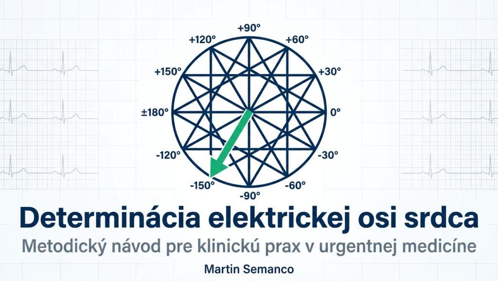
Abstract: Determining the heart’s electrical axis (the main QRS vector) is a basic diagnostic step in interpreting the 12-lead ECG. In emergency medicine, this parameter provides immediate insight into pathophysiological processes such as ventricular hypertrophy, intraventricular conduction disturbances, or acute right-heart strain. This article presents a methodical approach using geometric logic and visual mnemonics optimized for prehospital practice.
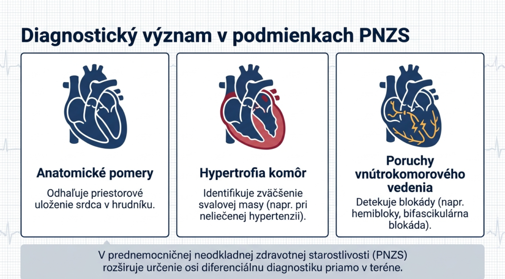
1. Clinical Significance in Prehospital Care
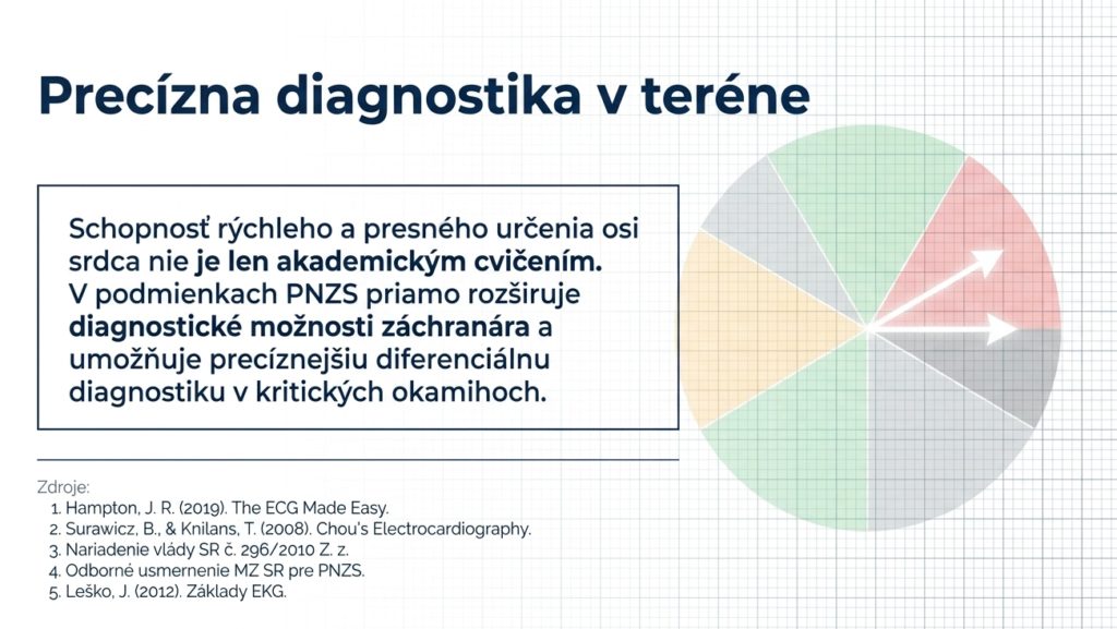
In the prehospital emergency care setting, analyzing the heart’s electrical axis is not merely a theoretical addendum. It represents a dynamic indicator that lets the field paramedic refine differential diagnosis in critically ill patients. Correctly interpreting the direction of the QRS vector reveals information about the heart’s anatomical position, identifies chronic changes (hypertrophy), and predicts the risk of sudden instability with conduction blocks.
2. Pathophysiological and Geometric Correlation
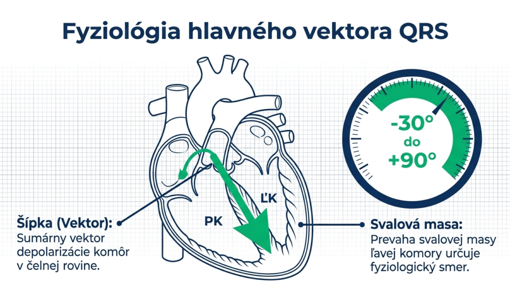
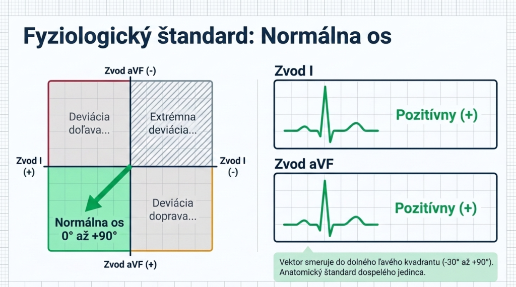
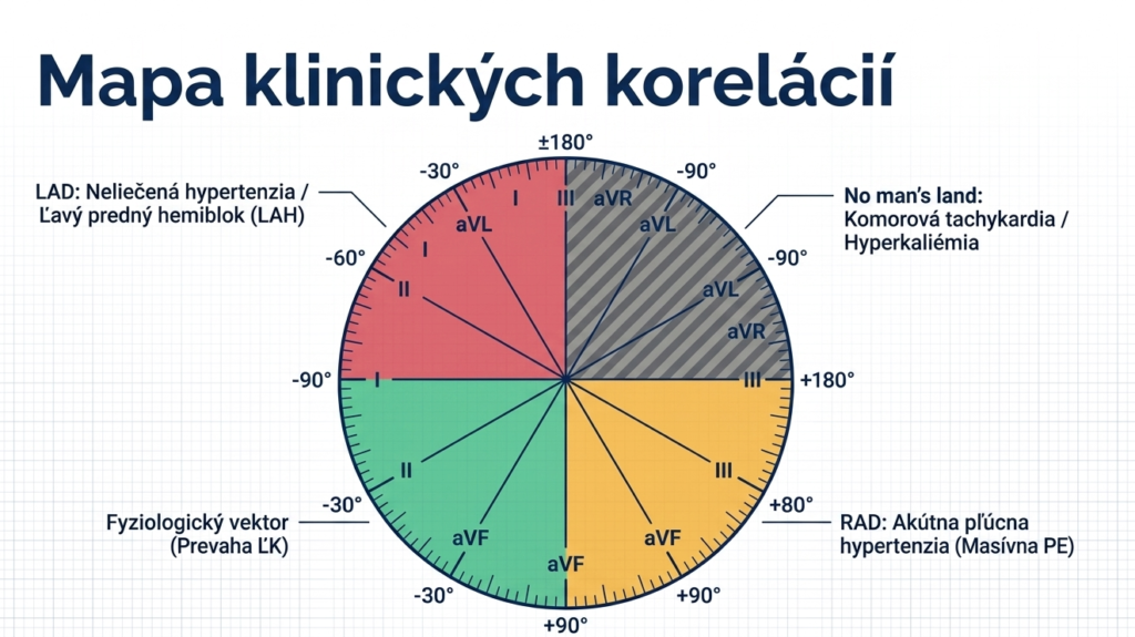
The heart’s electrical axis is defined as the summary vector of ventricular depolarization in the frontal plane. In a healthy adult, the left ventricle’s (LV) muscle mass dominates, directing the vector downward and to the left. The physiological axis range is -30 degrees to +90 degrees.
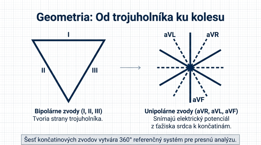
For analysis we use Bailey’s hexaxial reference system. This system integrates the bipolar limb leads (I, II, III), which form the sides of Einthoven’s triangle, with the unipolar augmented leads (aVR, aVL, aVF). The result is a 360-degree reference map, where each lead views the heart from a different angle.
3. Diagnostic Methodology: The Quadrant Method
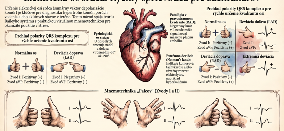
In emergency medicine practice, speed is emphasized. The most commonly used approach is the quadrant method. We assess QRS complex polarity (dominance of the positive or negative deflection) in two perpendicular leads: I and aVF.
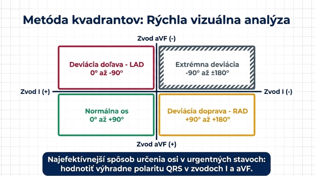
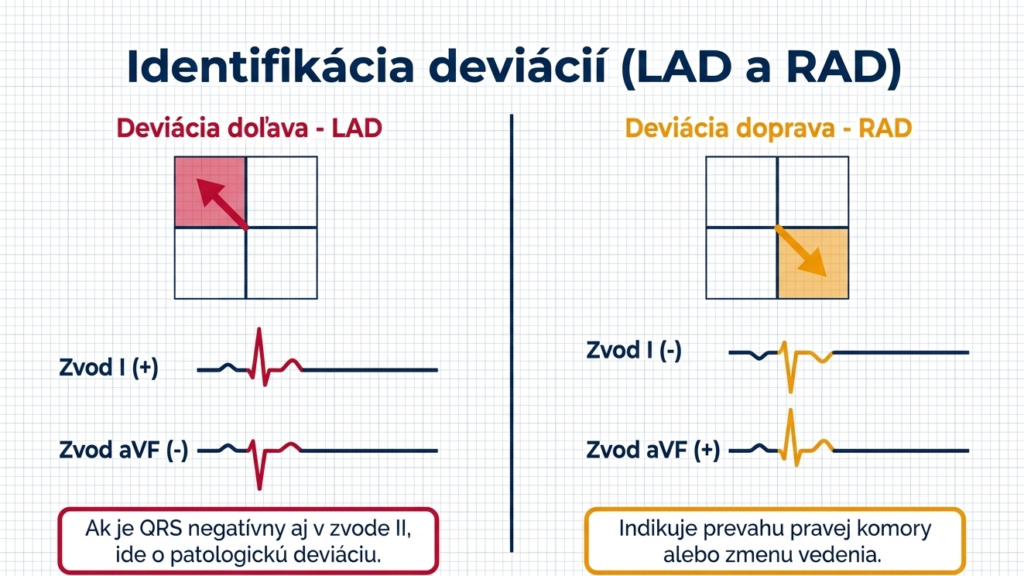
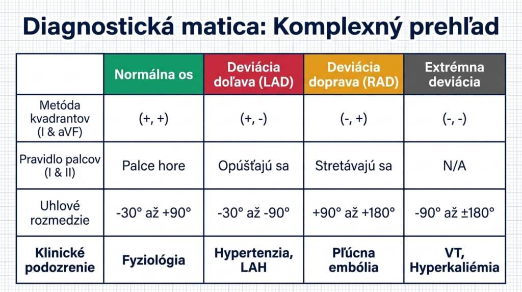
If the QRS is positive in both leads (I and aVF), we speak of a normal axis. Left deviation (LAD) manifests as positivity in I and negativity in aVF. If the trend is reversed (negative in I, positive in aVF), it’s right axis deviation (RAD). Extreme deviation (the so-called “no man’s land”) is present when both leads are negative.
4. Visual Mnemonic: The Thumbs-Up Rule
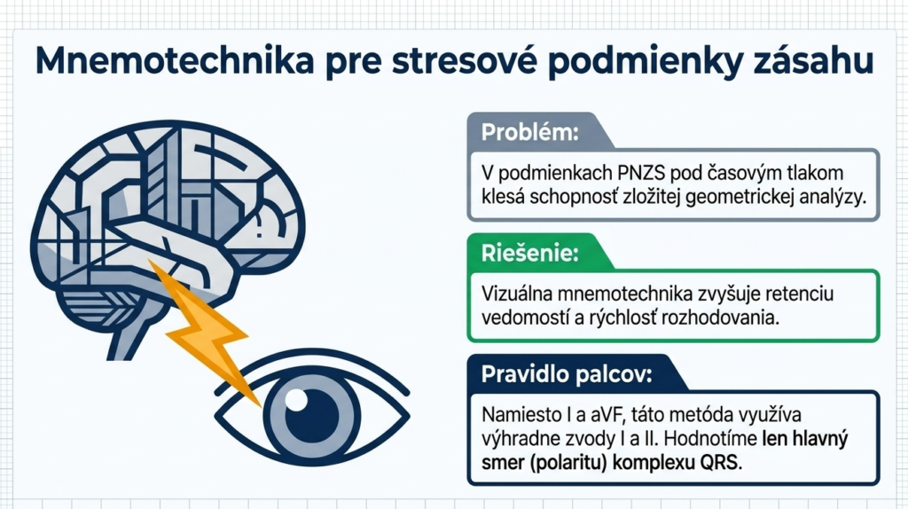
For working under the stressful conditions of a call, it’s ideal to use a mnemonic based on visual patterns. This method uses leads I and II.
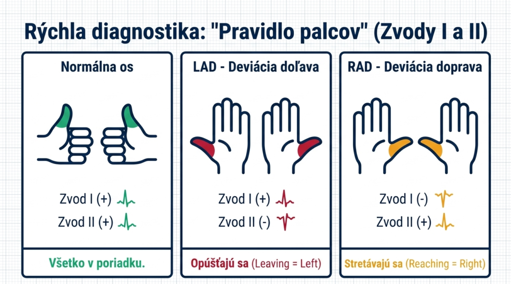
- Normal axis: Thumbs in both leads point up (both I and II are positive).
- LAD (Left Axis): Thumbs point away from each other (I up, II down) — mnemonic “Leaving” (moving apart).
- RAD (Right Axis): Thumbs point toward each other (I down, II up) — mnemonic “Reaching” (coming together).
5. Clinical Implications and Differential Diagnosis
Interpreting the finding must be correlated with the patient’s clinical state. The following overview summarizes the key pathophysiological correlations:
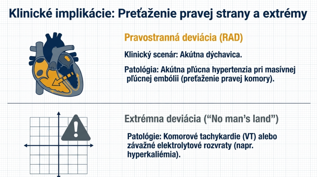
- Right axis deviation (RAD): Can signal pulmonary embolism, COPD, or acute right ventricular strain.
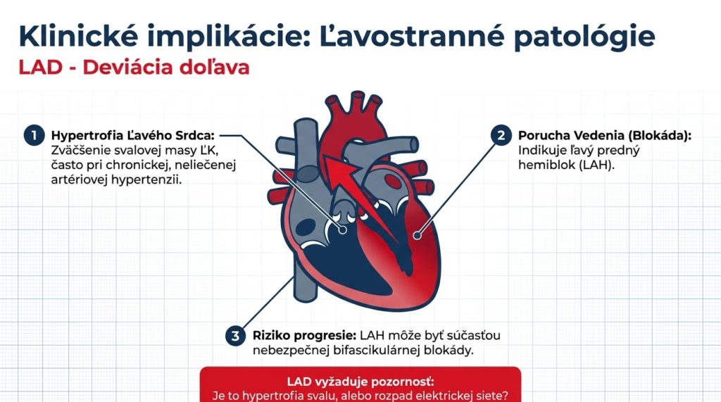
- Left axis deviation (LAD): Often accompanies left ventricular hypertrophy (in hypertension) or left anterior hemiblock (LAH), which can be part of a dangerous bifascicular block.
- Extreme deviation: A warning sign for ventricular tachycardia (VT) or critical hyperkalemia.
6. Conclusion
Determining the heart’s electrical axis is a key paramedic skill. Using mnemonic aids combined with a deep understanding of clinical correlations enables faster, more accurate diagnosis and subsequent treatment of the patient directly in the field.
References:
- Semanco, M. (2026): Determining the Heart’s Electrical Axis: A Methodical Guide for Clinical Practice.
- Hampton, J. R. (2019): The ECG Made Easy. 9th Ed., Elsevier.
- Leško, J. (2012): Fundamentals of ECG. Bratislava: Herba.
- Slovak Government Regulation No. 296/2010 Coll. on professional competence for the practice of healthcare professions.