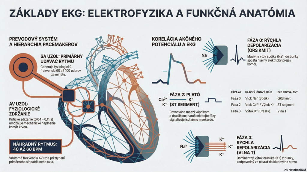
The Cardiac Conduction System (Specialized Cardiomyocytes) For adequate ECG interpretation, a paramedic must understand the hierarchy of pacemakers. The impulse originates in the sinoatrial (SA) node, which is the primary rhythm-setter (physiological rate 60 to 100 beats per minute).
1. Introduction: The Heart as a Network of Intelligent Batteries
Imagine every heart muscle cell as a miniature, high-performance rechargeable battery. At rest, this battery is “charged” to approximately -90 mV. We call this state the resting potential or polarization. It’s important to understand that this negative voltage isn’t just a static number, but accumulated potential energy ready for immediate work. Maintaining this charge is an active process requiring energy (ATP), while the “discharge” itself is a lightning-fast response to an electrical stimulus.
An ECG machine is nothing more than a sensitive array of “cameras” (electrodes) that monitor, from the body’s surface, the state these billions of batteries are currently in. When cells discharge (work) or recharge (rest), electrical differences arise that the ECG captures and draws as the familiar waveform.
This state of charged readiness lasts until the main control center issues the command for lightning-fast action.
——————————————————————————–
2. Phase 0 and the QRS Complex: A Rapid Energy Discharge
When the electrical impulse reaches the ventricles, phase 0 of the action potential begins — the moment the battery discharges. Fast sodium channels open, and a massive influx of sodium (Na+) into the cell occurs.
Depolarization: A lightning-fast change in charge from negative (-90 mV) to positive (approximately +20 mV). This rapid electrical discharge serves as the trigger for mechanical muscle contraction.
On the ECG, we see this event as the QRS complex. It’s the sharpest and most prominent part of the waveform, for two reasons:
- Extreme speed: Thanks to the His-Purkinje system, the impulse spreads through the ventricles at 2 to 4 m/s, ensuring nearly all ventricular cells discharge simultaneously. This is why a physiological QRS complex is very narrow — its duration is under 0.10 s.
- Mass: The ventricles represent the largest muscle mass, so their “discharge” generates the strongest electrical signal for our cameras.
Once the sodium storm subsides, the cell enters a mode of magical silence — the plateau phase.
——————————————————————————–
3. Phase 2 and the ST Segment: A Magical Balance (Plateau)
After the initial discharge, the heart needs a moment of stability. If the cell “relaxed” immediately after discharging, blood wouldn’t have time to be pumped through. That’s why phase 2 (plateau) occurs, during which a dynamic ionic balance is established in the cell.
| Ion Movement | ECG Result |
|---|---|
Dynamic balance: Ca2+ (in) ↔ K+ (out) | Isoelectric ST segment (flat line) |
The ST segment is flat on the ECG because net electrical current is zero at this moment. For a paramedic, however, this is a critical zone. Clinical significance: If cells are poorly oxygenated (ischemia), this ionic balance is disrupted, which the ECG camera immediately records as an ST-segment shift above or below the isoelectric line (elevation/depression). It’s a clear cry for help from the cells.
Once mechanical work (ejecting blood) is complete, the cell must perform a “major cleanup” to return to its original state.
——————————————————————————–
4. Phase 3 and the T Wave: Cleanup and Restart
Phase 3 is the process of rapid repolarization. Calcium channels close, and the cell sheds its positive charge by allowing potassium to flow massively outward. The battery “recharges” again to its original -90 mV.
This return to negativity is slower and more gradual than the sodium discharge, which defines the shape of the T wave:
- Key ion: Dominant potassium (K+) efflux.
- Electrical event: Return to resting polarization (repolarization).
- ECG shape: A broader, rounded T wave.
Without this energy-intensive restart, the heart would be unable to accept the next impulse, and electrical death would occur.
All these microscopic events combine into one perfectly synchronized whole, orchestrated by the conduction system.
——————————————————————————–
5. Synthesis: The Path of the Electrical Impulse from A to Z
Electricity travels from the SA node through the atria to the AV node. Here we encounter a phenomenon called decremental conduction — a physiological slowing of the impulse in the AV node (lasting 0.04 to 0.11 s). This delay is key: it gives the atria time to mechanically finish pushing blood into the ventricles before the ventricles themselves contract.
The following table serves as a complete diagnostic “cheat sheet”:
| ECG Wave/Segment | Action Potential Phase | Main Ion Movement | Biological Significance |
|---|---|---|---|
| P wave | Atrial depolarization | Na+ / Ca2+ influx | Atrial contraction |
| PR interval | Conduction (delay) | Ca2+ (slow channel) | Necessary pause to fill the ventricles |
| QRS complex | Phase 0 (Depolarization) | Rapid Sodium (Na+) influx | Massive ventricular contraction (discharge) |
| ST segment | Phase 2 (Plateau) | Ca2+ ↔ K+ balance | Sustaining contraction (work) |
| T wave | Phase 3 (Repolarization) | Potassium (K+) efflux | Electrical restart (recharging) |
Remember, whether a wave on the paper points up or down depends on the vector (direction) of electricity relative to your “camera.” If the wave travels toward the electrode, we see an upward deflection; if it travels away, a downward deflection.
——————————————————————————–
6. Final Summary for Quick Reference
For lightning-fast interpretation, link the ions to the phases in your mind:
- SODIUM (Na+) = START: Responsible for the fast QRS complex. Without it, there’s no action.
- CALCIUM (Ca2+) = WORK: Sustains the plateau phase (ST segment) and contraction strength.
- POTASSIUM (K+) = REST: Enables repolarization (T wave) and the battery’s return to standby mode.
——————————————————————————–
THE PARAMEDIC’S GOLDEN RULE: The ECG isn’t a direct picture of the mechanical pump, but a record of ionic currents. When you see pathology on the ST segment or T wave, you’re not just looking at a “line” — you’re looking at a cell fighting for its ionic balance and its ability to recharge.