Head and Neck Examination —
pupils, GCS, jugular veins, trachea, carotids
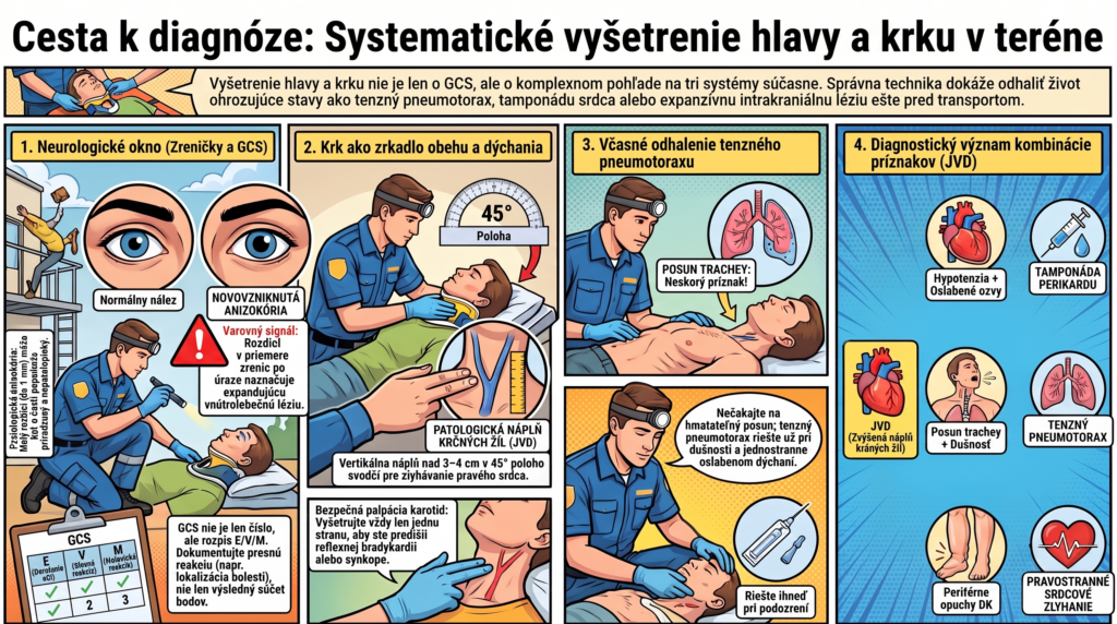
In the course of one quick exam, the head and neck provide data on three different systems — neurological, respiratory, and circulatory. Rarely is the concentration of diagnostically significant findings within a few square centimeters so high.
In the field, examining the head and neck is often reduced to “pupils plus GCS and let’s move on.” Yet this region contains jugular venous distension (a harbinger of right heart failure or tamponade), tracheal position (tension pneumothorax), carotid pulse symmetry, and many other findings that GCS alone doesn’t capture. This installment covers them systematically.
Pupils — more than just “equal, round, reactive”
Pupil examination is one of the few neurological findings that can be objectively assessed even in an unconscious patient, and therefore carries above-average weight in prehospital care.
Size — the normal range is approximately 2–5 mm, assessed under dim light before testing reactivity.
Symmetry (isocoria vs. anisocoria) — the diameter difference between the left and right pupil; new-onset anisocoria in a head-injury patient is a warning sign for an expanding intracranial lesion.
Light reactivity — direct (the illuminated pupil constricts) and consensual (the opposite pupil constricts simultaneously); assess the speed and extent of the reaction, not just its presence/absence.
Shape — an irregular shape (e.g., after eye surgery, after ocular trauma) must not be mistaken for a pathological neurological finding.
In a small percentage of the healthy population, mild physiological anisocoria (a difference up to about 1 mm) is present without any pathology. It’s therefore key to always ask a companion or family whether this is a longstanding condition for the patient — and document it. New-onset marked anisocoria is a different finding than lifelong mild asymmetry.
GCS — the components in practical execution
The Glasgow Coma Scale in the field is often reduced to a single number reported to the receiving facility, but its diagnostic value rests on how precisely the individual components are examined — not on the resulting sum.
| Component | How to examine correctly | Common mistake |
|---|---|---|
| Eye opening (E) | Gradual escalation: spontaneous → to voice → to a painful stimulus → none | Skipping the “to voice” step — going straight to a painful stimulus |
| Verbal response (V) | Orientation is assessed relative to person, place, time — not merely by the presence of speech | Rating “oriented” when the patient speaks fluently but doesn’t know where they are |
| Motor response (M) | A painful stimulus applied in a standardized way (above the supraorbital ridge or to the nail bed), the best response is scored | Mistaking non-targeted flexion (decortication) for targeted pain localization |
Jugular venous distension — an inconspicuous but fundamental finding
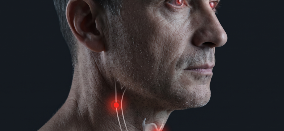
Distension of the external/internal jugular vein is one of the few physical findings that directly reflects the pressure conditions in the right atrium, and therefore the function of the right heart — without needing any device.
Right heart failure — the most common cause, often combined with peripheral edema.
Cardiac tamponade — combined with hypotension and muffled heart sounds, forms Beck’s triad.
Tension pneumothorax — combined with tracheal deviation and unilaterally diminished breath sounds.
Pulmonary embolism — acute right-heart strain in massive embolization.
Tracheal position
Palpating tracheal position in the suprasternal notch is a fast, simple finding with high clinical weight — its deviation from the midline is among the late but highly specific signs of tension pneumothorax.
By the time tracheal deviation becomes clinically palpable, tension pneumothorax is already at an advanced stage with significant circulatory impact. The absence of tracheal deviation therefore in no way rules out tension pneumothorax at an earlier stage — the diagnosis relies primarily on the combination of dyspnea, unilaterally diminished breath sounds, hyperresonant percussion, and circulatory instability, not on waiting for this late finding.
Carotid palpation
One side at a time — simultaneous bilateral carotid palpation can trigger reflex bradycardia or even syncope in sensitive patients (carotid sinus reflex).
Gentle pressure — excessive pressure on the carotid sinus can trigger the same reflex even with unilateral palpation.
Pulse symmetry — marked asymmetry may suggest stenosis or dissection, though in acute field decision-making this finding has only supplementary value.
Carotid palpation as a pulse-check substitute during CPR — the standard site for checking pulse in cardiac arrest in an adult; performed for a maximum of 10 seconds.
Three scenarios from practice
A patient after falling from a roof, GCS 9, new-onset anisocoria — the right pupil markedly wider, poorly reactive to light.
A patient with worsening dyspnea over several days, lower-extremity edema, jugular venous distension visible even while sitting.
A patient with a stab wound in the left subclavicular area, sudden worsening of breathing, tachycardia, dropping saturation.
What the next installment covers
Installment VYS-04 covers lung auscultation — stethoscope placement technique, the systematics of examination points, distinguishing individual breath sound phenomena (vesicular breathing, crackles, rhonchi, wheezes), and the limits of auscultation in a noisy ambulance environment.
- VYS-01 — Inspection, Palpation, Percussion, Auscultation: The Four Pillars of Physical Examination
- VYS-02 — Physical Examination in the Field vs. the Hospital
- VYS-03 — Head and Neck Examination (this article)
- VYS-04 — Lung Auscultation
- VYS-05 — Chest Examination
- VYS-06 — Heart Auscultation
- VYS-07 — Abdominal Examination
- VYS-08 — Peritoneal Signs and Acute Abdomen
- VYS-09 — Examination of the Limbs and Peripheral Circulation
- VYS-10 — Neurological Examination in the Field
- VYS-11 — The Skin as a Diagnostic Tool
- VYS-12 — Examining the Pediatric Patient
- VYS-13 — Examining the Geriatric Patient
- VYS-14 — From Examination to Decision
- Bickley LS, Szilagyi PG, Hoffman RM. Bates’ Guide to Physical Examination and History Taking. 13th ed. Wolters Kluwer, 2021.
- Teasdale G, Jennett B. Assessment of coma and impaired consciousness: a practical scale. The Lancet. 1974 — the original Glasgow Coma Scale publication.
- National Association of EMTs (NAEMT). PHTLS — Prehospital Trauma Life Support. 10th ed., 2023.
- American Heart Association. Guidelines for CPR and Emergency Cardiovascular Care, 2020 — carotid pulse assessment.
- Marx JA, et al. Rosen’s Emergency Medicine: Concepts and Clinical Practice. 10th ed. Elsevier, 2022 — chapter on tension pneumothorax and cardiac tamponade.