
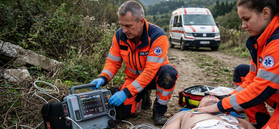
“The moment time stops in the back of the ambulance and the patient’s heart drops into a malignant arrhythmia, there’s no room to search through a menu. The LIFEPAK 15 isn’t a monitor — it’s your third crew member, and it has to respond to your touch with absolute certainty.”
Introduction: Why the Weight (Doesn’t) Matter
Many paramedics complain about the nine kilograms of “living iron” they have to haul up to the fourth floor. But the LIFEPAK 15 is built like a tank for one reason: survival. In an environment where devices fall off stretchers, get rained on, and where plastic viscosity changes in freezing cold, you need hardware that doesn’t know what an error message is. This article walks you through everything from mode settings to critical analysis of what you see on the display.
I. Dynamic Interpretation: 12-Lead ECG and SunVue
The LP15’s strength lies in diagnosing STEMI under pressure.
- SunVue mode: If you’re working in direct sunlight, switching to the black-and-white, high-contrast mode is the only reliable way to read ST elevations.
- Diagnostic filters: The LP15 features top-tier signal processing. On a shaking patient, the diagnostic filter (0.05–150 Hz) lets you see the pathology where other devices only draw a “sawtooth” line.
II. Pacing and Cardioversion: Surgical Precision of the Shock
- Transcutaneous pacing: In critical bradycardia, we’re looking for electrical and then mechanical “capture.” In Demand mode, the device “waits” for the patient’s own contraction, preventing dangerous R-on-T interference. Always confirm mechanical capture by palpating the right femoral artery.
- Synchronized cardioversion: In unstable SVT, the SYNC button is critical. The device marks each R wave with a small triangle.
- Caution: SYNC mode automatically switches off after every shock. If another shock is needed, you must reactivate it — otherwise you risk an asynchronous shock landing on the T wave and triggering fibrillation.
III. Capnometry: A Window into Metabolism and Ventilation
The integrated Oridion Microstream module is more valuable than the ECG itself.
- Resuscitation: A sudden jump in EtCO2 (for example, from 15 to 35 mmHg) is the fastest indicator of ROSC (return of spontaneous circulation).
- Obstruction: A waveform resembling a “shark fin” clearly indicates bronchospasm (asthma/COPD) even before auscultation.
IV. Artifact Analysis: Telling Truth from Noise
In the field, we treat the patient, not the monitor. But you still need to know when the technology is lying to you.
1. Motion artifact and muscle tremor
A chaotic line can resemble fine-wave ventricular fibrillation (VF).
- Verification: If the patient is talking to you, they are not in VF. Look for regular QRS complexes “showing through” the noise.
2. Mains interference (50 Hz)
Arises near power lines or when the ambulance’s metal frame contacts a live current. It appears as a “thick,” fuzzy isoelectric line and can mask small P waves or ST changes.
3. Wandering baseline artifact
The ECG trace “floats” up and down, making the ST segment impossible to read. The solution is better skin preparation and cable fixation so cables don’t tug on the electrodes with each breath.
4. CPR artifacts
Chest compressions generate spikes that correlate with the compression rate.
- Critical rule: Never assess the rhythm during compressions. A pause (max 5–10 s) is essential for a definitive shock decision.
V. Troubleshooting and Practical Tips
- SpCO and SpMet: In a patient from a fire, measure carboxyhemoglobin. The LP15 eliminates diagnostic uncertainty where a standard pulse oximeter falsely reads 100%.
- Paper failure: If the paper runs out during 12-lead printing, the device retains the recording in memory. After loading a new roll, you can print it from history.
- Pacer Detect: In patients with an implanted pacemaker, enable this function in the menu so the device highlights the spikes and doesn’t mistakenly count them as heart rate.
Conclusion: A Summary for Professionals
The LIFEPAK 15 is a comprehensive diagnostic terminal. Its ruggedness lets us focus on the patient, but it requires that we operate it automatically. If you’re ever in doubt about the rhythm, always follow this sequence: Check the patient – Compare with the clinical picture – Verify the technique (electrodes/cables).
Recommended Reading:
- Stryker / Physio-Control: LIFEPAK 15 Operating Instructions (2023).
- ERC Guidelines 2021: Advanced life support standards.
- Hovorka, J.: Arrhythmias in Emergency Medicine and Their Technical Management (2020).