This standard is based on the most current European Resuscitation Council (ERC) guidelines and is intended for professional paramedics and resuscitation team members working in both prehospital and hospital settings.
1. Recognizing cardiac arrest and basic procedures (BLS)
Early recognition of cardiac arrest and immediate action are the basic prerequisite for saving a life within the chain of survival.
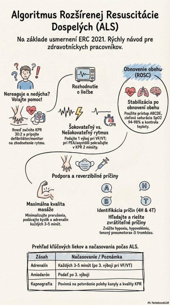
- Recognition: Assume cardiac arrest in any person who is unresponsive and not breathing, or not breathing normally. Slow, labored breathing (agonal breathing) is a key sign of cardiac arrest.
- A brief episode of seizure-like movements can occur at the onset of cardiac arrest. Reassess the patient once the episode ends; if they are not breathing, start resuscitation immediately.
- High-quality chest compressions: Start compressions as soon as possible. Compress the lower half of the sternum to a depth of at least 5 cm but no more than 6 cm, at a rate of 100–120/min.
- After each compression, allow full chest recoil and do not lean on the chest; minimize any interruptions.
- Perform resuscitation at a ratio of 30 compressions to 2 rescue breaths.
2. Advanced life support (ALS)
High-quality chest compressions with minimal interruptions and early defibrillation remain the foundation of successful ALS.
A. Shockable rhythms (ventricular fibrillation – VF / pulseless ventricular tachycardia – pVT)
- Defibrillate as quickly as possible (first shock energy of at least 150 J for a biphasic shock, or 120–150 J).
- Minimize interruptions: Pause compressions only immediately before delivering the shock, aiming for a pause of less than 5 seconds. Resume CPR immediately after the shock for 2 minutes.
- Pharmacotherapy: If a shockable rhythm persists, give epinephrine 1 mg IV/IO and amiodarone 300 mg IV/IO immediately after the 3rd shock. Repeat epinephrine (1 mg) every 3–5 minutes thereafter.
B. Non-shockable rhythms (asystole / pulseless electrical activity – PEA)
- Continue CPR immediately in 2-minute cycles.
- Give epinephrine 1 mg IV/IO as soon as possible. Repeat every 3–5 minutes thereafter.
3. Airway management and ventilation
- Proceed according to the rescuer’s skill level. If needed, start with basic techniques and escalate progressively.
- Tracheal intubation should only be performed by rescuers with a high success rate (defined as greater than 95% success within two attempts). Do not interrupt chest compressions for intubation for more than 5 seconds.
- To confirm correct tracheal tube placement, always use capnography (the EtCO2 waveform), which also lets you monitor CPR quality.
- Once the airway is secured with a tracheal tube or a supraglottic device, ventilate the patient continuously at a rate of 10 breaths per minute and do not interrupt chest compressions for breaths. Each breath should take 1 second, delivered until visible chest rise; give patients the highest available fraction of inspired oxygen.
4. Treating reversible causes (the 4 Hs and 4 Ts)
Throughout resuscitation, always search for reversible causes of the arrest and address them immediately.
- 4 Hs: Hypoxia, Hypovolemia, Hypo-/Hyperkalemia and other metabolic disorders, Hypo-/Hyperthermia.
- 4 Ts: Thrombosis (coronary or pulmonary), Tension pneumothorax, cardiac Tamponade, Toxins.
- Point-of-care ultrasound (POCUS): Experienced rescuers should use it to diagnose treatable causes such as tamponade or pneumothorax. However, the exam must not cause an additional or prolonged interruption in chest compressions.
5. Traumatic cardiac arrest (TCA)
Procedures for TCA differ fundamentally. While compressions dominate in cardiac causes, in trauma simultaneous treatment of reversible causes takes priority over chest compressions.
- Key interventions are: immediate control of catastrophic external hemorrhage (e.g., tourniquets, hemostatic dressings), airway management with maximal oxygenation, bilateral chest decompression (thoracostomy/thoracocentesis), release of tamponade, and pelvic binding.
- Chest compressions must not delay these life-saving interventions; rapid fluid resuscitation, ideally with blood products, is also essential.
6. Post-resuscitation care (after ROSC)
Care begins immediately after return of spontaneous circulation (ROSC), using an ABCDE approach.
- Airway and breathing: Titrate inspired oxygen (FiO2) to achieve an SpO2 of 94–98%, and ventilate the lungs to a state of normocapnia. Strictly avoid both hypoxemia and hyperoxemia.
- Circulation: Obtain a 12-lead ECG. If the patient is not hypotensive, aim to maintain normovolemia. Avoid hypotension and maintain a target systolic blood pressure > 100 mmHg (using fluids or vasopressors as needed). With ST-elevation on the ECG (STEMI), direct the patient straight to emergency coronary angiography/PCI.
- Temperature control: For comatose patients after ROSC, use targeted temperature management (TTM). Maintain a constant temperature of 32–36°C for at least 24 hours, and strictly prevent fever for at least 72 hours.